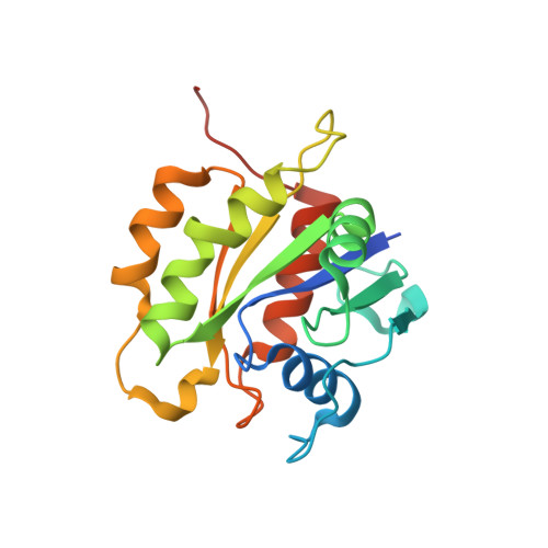
6D71

Crystal Structure of the Human Miro1 N-terminal GTPase bound to GTP


Experimental Data Snapshot

  • Method: X-RAY DIFFRACTION
  • Resolution: 1.72 Å
  • R-Value Free: 0.220 
  • R-Value Work: 0.182 
  • R-Value Observed: 0.184 

wwPDB Validation   3D Report Full Report


Ligand Structure Quality Assessment 


This is version 1.2 of the entry. See complete history


Literature

Insight into human Miro1/2 domain organization based on the structure of its N-terminal GTPase.

Smith, K.P.Focia, P.J.Chakravarthy, S.Landahl, E.C.Klosowiak, J.L.Rice, S.E.Freymann, D.M.

(2020) J Struct Biol 212: 107656-107656

  • DOI: https://doi.org/10.1016/j.jsb.2020.107656
  • Primary Citation of Related Structures:  
    6D71

  • PubMed Abstract: 

    Dysfunction in mitochondrial dynamics is believed to contribute to a host of neurological disorders and has recently been implicated in cancer metastasis. The outer mitochondrial membrane adapter protein Miro functions in the regulation of mitochondrial mobility and degradation, however, the structural basis for its roles in mitochondrial regulation remain unknown. Here, we report a 1.7Å crystal structure of N-terminal GTPase domain (nGTPase) of human Miro1 bound unexpectedly to GTP, thereby revealing a non-catalytic configuration of the putative GTPase active site. We identify two conserved surfaces of the nGTPase, the "SELFYY" and "ITIP" motifs, that are potentially positioned to mediate dimerization or interaction with binding partners. Additionally, we report small angle X-ray scattering (SAXS) data obtained from the intact soluble HsMiro1 and its paralog HsMiro2. Taken together, the data allow modeling of a crescent-shaped assembly of the soluble domain of HsMiro1/2. PDB RSEFERENCE: Crystal structure of the human Miro1 N-terminal GTPase bound to GTP, 6D71.


  • Organizational Affiliation

    Department of Cell & Molecular Biology, Feinberg School of Medicine, Northwestern University, 303 East Chicago Avenue, Chicago, IL 60611, USA. Electronic address: kyle.smith.phd@gmail.com.


Macromolecules
Find similar proteins by:  (by identity cutoff)  |  3D Structure
Entity ID: 1
MoleculeChains Sequence LengthOrganismDetailsImage
Mitochondrial Rho GTPase 1
A, B
186Homo sapiensMutation(s): 0 
Gene Names: RHOT1ARHT1
EC: 3.6.5
UniProt & NIH Common Fund Data Resources
Find proteins for Q8IXI2 (Homo sapiens)
Explore Q8IXI2 
Go to UniProtKB:  Q8IXI2
PHAROS:  Q8IXI2
GTEx:  ENSG00000126858 
Entity Groups  
Sequence Clusters30% Identity50% Identity70% Identity90% Identity95% Identity100% Identity
UniProt GroupQ8IXI2
Sequence Annotations
Expand
  • Reference Sequence
Experimental Data & Validation

Experimental Data

  • Method: X-RAY DIFFRACTION
  • Resolution: 1.72 Å
  • R-Value Free: 0.220 
  • R-Value Work: 0.182 
  • R-Value Observed: 0.184 
  • Space Group: P 21 21 21
Unit Cell:
Length ( Å )Angle ( ˚ )
a = 41.35α = 90
b = 78.27β = 90
c = 99.78γ = 90
Software Package:
Software NamePurpose
MOSFLMdata reduction
Aimlessdata scaling
PHENIXphasing
PHENIXrefinement

Structure Validation

View Full Validation Report


Ligand Structure Quality Assessment 


Entry History & Funding Information

Deposition Data


Funding OrganizationLocationGrant Number
National Institutes of Health/National Human Genome Research Institute (NIH/NHGRI)United StatesR01GM107209
National Institutes of Health/National Human Genome Research Institute (NIH/NHGRI)United StatesT32GM008382
National Institutes of Health/National Cancer Institute (NIH/NCI)United StatesNCI-CCSG-P30-CA060553

Revision History  (Full details and data files)

  • Version 1.0: 2019-10-09
    Type: Initial release
  • Version 1.1: 2019-12-04
    Changes: Author supporting evidence
  • Version 1.2: 2022-10-05
    Changes: Advisory, Database references, Derived calculations