6D4F

Crystal structure of PTP epsilon D2 domain (A455N/V457Y/E597D)


Experimental Data Snapshot

  • Method: X-RAY DIFFRACTION
  • Resolution: 1.91 Å
  • R-Value Free: 0.225 
  • R-Value Work: 0.190 
  • R-Value Observed: 0.193 

wwPDB Validation   3D Report Full Report


This is version 1.1 of the entry. See complete history


Literature

High-resolution crystal structures of the D1 and D2 domains of protein tyrosine phosphatase epsilon for structure-based drug design.

Lountos, G.T.Raran-Kurussi, S.Zhao, B.M.Dyas, B.K.Burke Jr., T.R.Ulrich, R.G.Waugh, D.S.

(2018) Acta Crystallogr D Struct Biol 74: 1015-1026

  • DOI: https://doi.org/10.1107/S2059798318011919
  • Primary Citation of Related Structures:  
    6D3F, 6D4D, 6D4F

  • PubMed Abstract: 

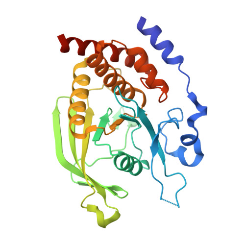
    Here, new crystal structures are presented of the isolated membrane-proximal D1 and distal D2 domains of protein tyrosine phosphatase epsilon (PTPℇ), a protein tyrosine phosphatase that has been shown to play a positive role in the survival of human breast cancer cells. A triple mutant of the PTPℇ D2 domain (A455N/V457Y/E597D) was also constructed to reconstitute the residues of the PTPℇ D1 catalytic domain that are important for phosphatase activity, resulting in only a slight increase in the phosphatase activity compared with the native D2 protein. The structures reported here are of sufficient resolution for structure-based drug design, and a microarray-based assay for high-throughput screening to identify small-molecule inhibitors of the PTPℇ D1 domain is also described.


  • Organizational Affiliation

    Basic Science Program, Frederick National Laboratory for Cancer Research sponsored by the National Cancer Institute, Frederick, MD 21702, USA.


Macromolecules
Find similar proteins by:  (by identity cutoff)  |  3D Structure
Entity ID: 1
MoleculeChains Sequence LengthOrganismDetailsImage
Receptor-type tyrosine-protein phosphatase epsilon279Homo sapiensMutation(s): 3 
Gene Names: PTPRE
EC: 3.1.3.48
UniProt & NIH Common Fund Data Resources
Find proteins for P23469 (Homo sapiens)
Explore P23469 
Go to UniProtKB:  P23469
PHAROS:  P23469
GTEx:  ENSG00000132334 
Entity Groups  
Sequence Clusters30% Identity50% Identity70% Identity90% Identity95% Identity100% Identity
UniProt GroupP23469
Sequence Annotations
Expand
  • Reference Sequence
Small Molecules
Ligands 1 Unique
IDChains Name / Formula / InChI Key2D Diagram3D Interactions
1PE
Query on 1PE

Download Ideal Coordinates CCD File 
B [auth A]PENTAETHYLENE GLYCOL
C10 H22 O6
JLFNLZLINWHATN-UHFFFAOYSA-N
Experimental Data & Validation

Experimental Data

  • Method: X-RAY DIFFRACTION
  • Resolution: 1.91 Å
  • R-Value Free: 0.225 
  • R-Value Work: 0.190 
  • R-Value Observed: 0.193 
  • Space Group: P 21 21 21
Unit Cell:
Length ( Å )Angle ( ˚ )
a = 61.47α = 90
b = 73.489β = 90
c = 75.656γ = 90
Software Package:
Software NamePurpose
PHENIXrefinement
HKL-3000data reduction
HKL-3000data scaling
PHASERphasing

Structure Validation

View Full Validation Report


Entry History 

Deposition Data

Revision History  (Full details and data files)

  • Version 1.0: 2018-10-17
    Type: Initial release
  • Version 1.1: 2023-10-04
    Changes: Data collection, Database references, Refinement description