6D1R

Structure of Staphylococcus aureus RNase P protein at 2.0 angstrom


Experimental Data Snapshot

  • Method: X-RAY DIFFRACTION
  • Resolution: 2.00 Å
  • R-Value Free: 0.234 
  • R-Value Work: 0.214 
  • R-Value Observed: 0.216 

wwPDB Validation   3D Report Full Report


This is version 1.2 of the entry. See complete history


Literature

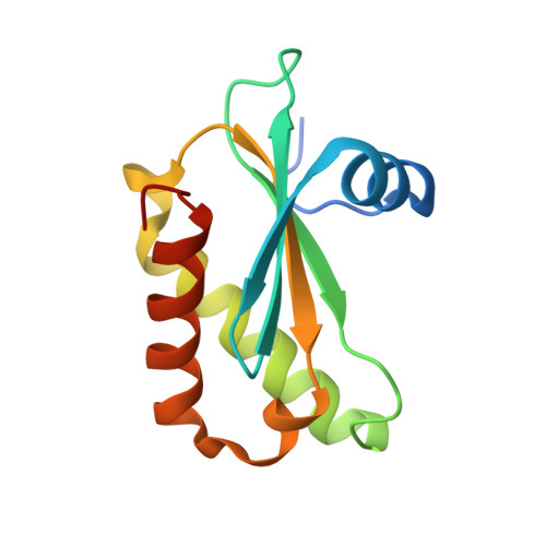
Crystal structure of the ribonuclease-P-protein subunit from Staphylococcus aureus.

Ha, L.Colquhoun, J.Noinaj, N.Das, C.Dunman, P.M.Flaherty, D.P.

(2018) Acta Crystallogr F Struct Biol Commun 74: 632-637

  • DOI: https://doi.org/10.1107/S2053230X18011512
  • Primary Citation of Related Structures:  
    6D1R

  • PubMed Abstract: 

    Staphylococcus aureus ribonuclease-P-protein subunit (RnpA) is a promising antimicrobial target that is a key protein component for two essential cellular processes, RNA degradation and transfer-RNA (tRNA) maturation. The first crystal structure of RnpA from the pathogenic bacterial species, S. aureus, is reported at 2.0 Å resolution. The structure presented maintains key similarities with previously reported RnpA structures from bacteria and archaea, including the highly conserved RNR-box region and aromatic residues in the precursor-tRNA 5'-leader-binding domain. This structure will be instrumental in the pursuit of structure-based designed inhibitors targeting RnpA-mediated RNA processing as a novel therapeutic approach for treating S. aureus infections.


  • Organizational Affiliation

    Department of Medicinal Chemistry and Molecular Pharmacology, College of Pharmacy, Purdue University, West Lafayette, IN 47907, USA.


Macromolecules
Find similar proteins by:  (by identity cutoff)  |  3D Structure
Entity ID: 1
MoleculeChains Sequence LengthOrganismDetailsImage
Ribonuclease P protein component119Staphylococcus aureusMutation(s): 0 
Gene Names: rnpA
EC: 3.1.26.5
UniProt
Find proteins for P0A0H5 (Staphylococcus aureus)
Explore P0A0H5 
Go to UniProtKB:  P0A0H5
Entity Groups  
Sequence Clusters30% Identity50% Identity70% Identity90% Identity95% Identity100% Identity
UniProt GroupP0A0H5
Sequence Annotations
Expand
  • Reference Sequence
Experimental Data & Validation

Experimental Data

  • Method: X-RAY DIFFRACTION
  • Resolution: 2.00 Å
  • R-Value Free: 0.234 
  • R-Value Work: 0.214 
  • R-Value Observed: 0.216 
  • Space Group: P 3 2 1
Unit Cell:
Length ( Å )Angle ( ˚ )
a = 72.159α = 90
b = 72.159β = 90
c = 58.958γ = 120
Software Package:
Software NamePurpose
PHENIXrefinement
Aimlessdata scaling
PHASERphasing
PDB_EXTRACTdata extraction
HKL-2000data reduction

Structure Validation

View Full Validation Report


Entry History 

Deposition Data

Revision History  (Full details and data files)

  • Version 1.0: 2018-09-26
    Type: Initial release
  • Version 1.1: 2018-10-17
    Changes: Data collection, Database references
  • Version 1.2: 2024-03-13
    Changes: Data collection, Database references