6COJ

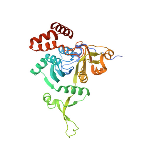
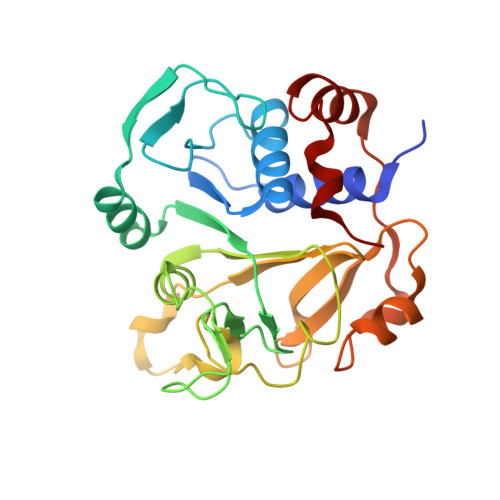
Crystal structure of Rhodococcus jostii RHA1 IpdAB E105A COCHEA-COA complex


Experimental Data Snapshot

  • Method: X-RAY DIFFRACTION
  • Resolution: 1.40 Å
  • R-Value Free: 0.173 
  • R-Value Work: 0.152 
  • R-Value Observed: 0.153 

wwPDB Validation   3D Report Full Report


Ligand Structure Quality Assessment 


This is version 1.3 of the entry. See complete history


Literature

IpdAB, a virulence factor inMycobacterium tuberculosis, is a cholesterol ring-cleaving hydrolase.

Crowe, A.M.Workman, S.D.Watanabe, N.Worrall, L.J.Strynadka, N.C.J.Eltis, L.D.

(2018) Proc Natl Acad Sci U S A 115: E3378-E3387

  • DOI: https://doi.org/10.1073/pnas.1717015115
  • Primary Citation of Related Structures:  
    6CO6, 6CO9, 6COJ, 6CON

  • PubMed Abstract: 
  • Mycobacterium tuberculosis ( Mtb ) grows on host-derived cholesterol during infection. IpdAB, found in all steroid-degrading bacteria and a determinant of pathogenicity, has been implicated in the hydrolysis of the last steroid ring. Phylogenetic analyses revealed that IpdAB orthologs form a clade of CoA transferases (CoTs) ...

    Mycobacterium tuberculosis ( Mtb ) grows on host-derived cholesterol during infection. IpdAB, found in all steroid-degrading bacteria and a determinant of pathogenicity, has been implicated in the hydrolysis of the last steroid ring. Phylogenetic analyses revealed that IpdAB orthologs form a clade of CoA transferases (CoTs). In a coupled assay with a thiolase, IpdAB transformed the cholesterol catabolite ( R )-2-(2-carboxyethyl)-3-methyl-6-oxocyclohex-1-ene-1-carboxyl-CoA (COCHEA-CoA) and CoASH to 4-methyl-5-oxo-octanedioyl-CoA (MOODA-CoA) and acetyl-CoA with high specificity ( k cat / K m = 5.8 ± 0.8 × 10 4 M -1 ⋅s -1 ). The structure of MOODA-CoA was consistent with IpdAB hydrolyzing COCHEA-CoA to a β-keto-thioester, a thiolase substrate. Contrary to characterized CoTs, IpdAB exhibited no activity toward small CoA thioesters. Further, IpdAB lacks the catalytic glutamate residue that is conserved in the β-subunit of characterized CoTs and a glutamyl-CoA intermediate was not trapped during turnover. By contrast, Glu105 A , conserved in the α-subunit of IpdAB, was essential for catalysis. A crystal structure of the IpdAB·COCHEA-CoA complex, solved to 1.4 Å, revealed that Glu105 A is positioned to act as a catalytic base. Upon titration with COCHEA-CoA, the E105A A variant accumulated a yellow-colored species (λ max = 310 nm; K d = 0.4 ± 0.2 μM) typical of β-keto enolates. In the presence of D 2 O, IpdAB catalyzed the deuteration of COCHEA-CoA adjacent to the hydroxylation site at rates consistent with k cat Based on these data and additional IpdAB variants, we propose a retro-Claisen condensation-like mechanism for the IpdAB-mediated hydrolysis of COCHEA-CoA. This study expands the range of known reactions catalyzed by the CoT superfamily and provides mechanistic insight into an important determinant of Mtb pathogenesis.


    Organizational Affiliation

    Department of Microbiology and Immunology, Life Sciences Institute, The University of British Columbia, Vancouver, BC, Canada, V6T 1Z3.


Macromolecules
Find similar proteins by:  (by identity cutoff)  |  3D Structure
Entity ID: 1
MoleculeChainsSequence LengthOrganismDetailsImage
Probable CoA-transferase alpha subunitA308Rhodococcus jostii RHA1Mutation(s): 1 
Gene Names: RHA1_ro04651ipdA
EC: 4.1.99
UniProt
Find proteins for Q0S7P9 (Rhodococcus jostii (strain RHA1))
Explore Q0S7P9 
Go to UniProtKB:  Q0S7P9
Entity Groups  
Sequence Clusters30% Identity50% Identity70% Identity90% Identity95% Identity100% Identity
UniProt GroupQ0S7P9
Protein Feature View
Expand
  • Reference Sequence
Find similar proteins by:  (by identity cutoff)  |  3D Structure
Entity ID: 2
MoleculeChainsSequence LengthOrganismDetailsImage
Probable CoA-transferase beta subunitB253Rhodococcus jostii RHA1Mutation(s): 0 
Gene Names: RHA1_ro04650ipdB
EC: 4.1.99
UniProt
Find proteins for Q0S7Q0 (Rhodococcus jostii (strain RHA1))
Explore Q0S7Q0 
Go to UniProtKB:  Q0S7Q0
Entity Groups  
Sequence Clusters30% Identity50% Identity70% Identity90% Identity95% Identity100% Identity
UniProt GroupQ0S7Q0
Protein Feature View
Expand
  • Reference Sequence
Small Molecules
Ligands 2 Unique
IDChainsName / Formula / InChI Key2D Diagram3D Interactions
F8G (Subject of Investigation/LOI)
Query on F8G

Download Ideal Coordinates CCD File 
E [auth B]S-{(3R,5R,9R)-1-[(2R,3S,4R,5R)-5-(6-amino-9H-purin-9-yl)-4-hydroxy-3-(phosphonooxy)tetrahydrofuran-2-yl]-3,5,9-trihydroxy-8,8-dimethyl-3,5-dioxido-10,14-dioxo-2,4,6-trioxa-11,15-diaza-3lambda~5~,5lambda~5~-diphosphaheptadecan-17-yl} (5R,10R)-7-hydroxy-10-methyl-2-oxo-1-oxaspiro[4.5]dec-6-ene-6-carbothioate (non-preferred name)
C32 H48 N7 O20 P3 S
COMHLEMSBYWTLA-LJKJHTHQSA-N
 Ligand Interaction
SO4
Query on SO4

Download Ideal Coordinates CCD File 
C [auth A],
D [auth A],
F [auth B]
SULFATE ION
O4 S
QAOWNCQODCNURD-UHFFFAOYSA-L
 Ligand Interaction
Experimental Data & Validation

Experimental Data

  • Method: X-RAY DIFFRACTION
  • Resolution: 1.40 Å
  • R-Value Free: 0.173 
  • R-Value Work: 0.152 
  • R-Value Observed: 0.153 
  • Space Group: P 43 21 2
Unit Cell:
Length ( Å )Angle ( ˚ )
a = 69.172α = 90
b = 69.172β = 90
c = 241.873γ = 90
Software Package:
Software NamePurpose
PHENIXrefinement
Aimlessdata scaling
PDB_EXTRACTdata extraction
XDSdata reduction
PHASERphasing

Structure Validation

View Full Validation Report


Ligand Structure Quality Assessment 


Entry History 

Deposition Data

Revision History  (Full details and data files)

  • Version 1.0: 2018-03-28
    Type: Initial release
  • Version 1.1: 2018-04-11
    Changes: Data collection, Database references
  • Version 1.2: 2018-04-25
    Changes: Data collection, Database references
  • Version 1.3: 2018-06-13
    Changes: Data collection, Structure summary