6CD2

Crystal structure of the PapC usher bound to the chaperone-adhesin PapD-PapG


Experimental Data Snapshot

  • Method: X-RAY DIFFRACTION
  • Resolution: 3.70 Å
  • R-Value Free: 0.329 
  • R-Value Work: 0.282 
  • R-Value Observed: 0.284 

wwPDB Validation   3D Report Full Report


This is version 1.4 of the entry. See complete history


Literature

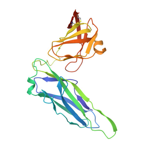
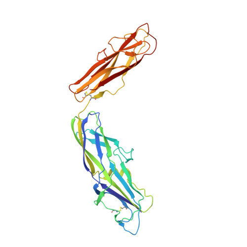
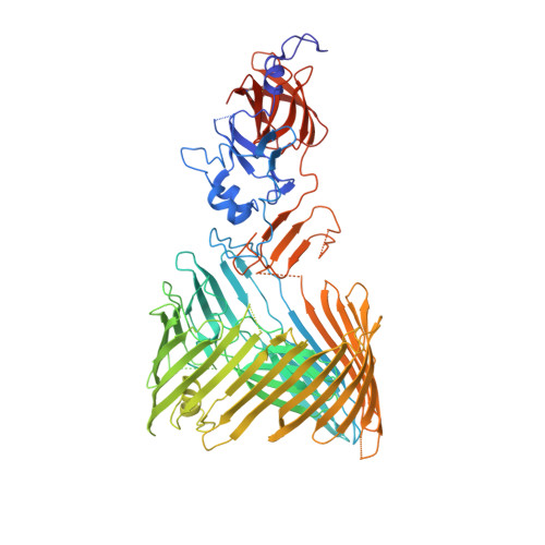
Structural basis for usher activation and intramolecular subunit transfer in P pilus biogenesis in Escherichia coli.

Omattage, N.S.Deng, Z.Pinkner, J.S.Dodson, K.W.Almqvist, F.Yuan, P.Hultgren, S.J.

(2018) Nat Microbiol 3: 1362-1368

  • DOI: https://doi.org/10.1038/s41564-018-0255-y
  • Primary Citation of Related Structures:  
    6CD2

  • PubMed Abstract: 

    Chaperone-usher pathway pili are extracellular proteinaceous fibres ubiquitously found on Gram-negative bacteria, and mediate host-pathogen interactions and biofilm formation critical in pathogenesis in numerous human diseases 1 . During pilus assembly, an outer membrane macromolecular machine called the usher catalyses pilus biogenesis from the individual subunits that are delivered as chaperone-subunit complexes in the periplasm. The usher orchestrates pilus assembly using all five functional domains: a 24-stranded transmembrane β-barrel translocation domain, a β-sandwich plug domain, an amino-terminal periplasmic domain and two carboxy-terminal periplasmic domains (CTD1 and CTD2) 2-6 . Despite extensive structural and functional characterization, the mechanism by which the usher is activated to initiate pilus biogenesis is unknown. Here, we present the crystal structure of the full-length PapC usher from Escherichia coli in complex with its cognate PapDG chaperone-subunit complex in a pre-activation state, elucidating molecular details of how the usher is specifically engaged by allosteric interactions with its substrate preceding activation and how the usher facilitates the transfer of subunits from the amino-terminal periplasmic domain to the CTDs during pilus assembly. This work elucidates the intricate workings of a molecular machine that catalyses chaperone-usher pathway pilus assembly and opens the door for the development of potent inhibitors to block pilus biogenesis.


  • Organizational Affiliation

    Department of Molecular Microbiology, Washington University in St Louis, St Louis, MO, USA.


Macromolecules
Find similar proteins by:  (by identity cutoff)  |  3D Structure
Entity ID: 1
MoleculeChains Sequence LengthOrganismDetailsImage
Chaperone protein PapD221Escherichia coliMutation(s): 0 
Gene Names: papD
Membrane Entity: Yes 
UniProt
Find proteins for P15319 (Escherichia coli)
Explore P15319 
Go to UniProtKB:  P15319
Entity Groups  
Sequence Clusters30% Identity50% Identity70% Identity90% Identity95% Identity100% Identity
UniProt GroupP15319
Sequence Annotations
Expand
  • Reference Sequence
Find similar proteins by:  (by identity cutoff)  |  3D Structure
Entity ID: 2
MoleculeChains Sequence LengthOrganismDetailsImage
PapGII adhesin protein316Escherichia coliMutation(s): 0 
Membrane Entity: Yes 
UniProt
Find proteins for Q47450 (Escherichia coli)
Explore Q47450 
Go to UniProtKB:  Q47450
Entity Groups  
Sequence Clusters30% Identity50% Identity70% Identity90% Identity95% Identity100% Identity
UniProt GroupQ47450
Sequence Annotations
Expand
  • Reference Sequence
Find similar proteins by:  (by identity cutoff)  |  3D Structure
Entity ID: 3
MoleculeChains Sequence LengthOrganismDetailsImage
Outer membrane usher protein PapC757Escherichia coliMutation(s): 0 
Gene Names: PapC
Membrane Entity: Yes 
UniProt
Find proteins for P07110 (Escherichia coli)
Explore P07110 
Go to UniProtKB:  P07110
Entity Groups  
Sequence Clusters30% Identity50% Identity70% Identity90% Identity95% Identity100% Identity
UniProt GroupP07110
Sequence Annotations
Expand
  • Reference Sequence
Experimental Data & Validation

Experimental Data

  • Method: X-RAY DIFFRACTION
  • Resolution: 3.70 Å
  • R-Value Free: 0.329 
  • R-Value Work: 0.282 
  • R-Value Observed: 0.284 
  • Space Group: C 2 2 21
Unit Cell:
Length ( Å )Angle ( ˚ )
a = 204.303α = 90
b = 300.71β = 90
c = 100.515γ = 90
Software Package:
Software NamePurpose
REFMACrefinement
HKL-2000data reduction
HKL-2000data scaling
PHASERphasing

Structure Validation

View Full Validation Report


Entry History & Funding Information

Deposition Data


Funding OrganizationLocationGrant Number
National Institutes of Health/National Institute Of Allergy and Infectious Diseases (NIH/NIAID)United StatesR01AI029549
National Institutes of Health/National Institute Of Allergy and Infectious Diseases (NIH/NIAID)United StatesR01AI048689
National Science Foundation (NSF, United States)United StatesDGE-1745038

Revision History  (Full details and data files)

  • Version 1.0: 2018-10-03
    Type: Initial release
  • Version 1.1: 2018-10-31
    Changes: Data collection, Database references
  • Version 1.2: 2018-12-12
    Changes: Data collection, Database references
  • Version 1.3: 2019-11-27
    Changes: Author supporting evidence
  • Version 1.4: 2023-10-04
    Changes: Data collection, Database references, Refinement description