6C2C | pdb_00006c2c

The molecular basis for the functional evolution of an organophosphate hydrolysing enzyme


Experimental Data Snapshot

  • Method: X-RAY DIFFRACTION
  • Resolution: 1.60 Å
  • R-Value Free: 
    0.204 (Depositor), 0.205 (DCC) 
  • R-Value Work: 
    0.173 (Depositor), 0.175 (DCC) 
  • R-Value Observed: 
    0.175 (Depositor) 

wwPDB Validation 3D Report Full Report

Validation slider image for 6C2C

This is version 1.3 of the entry. See complete history

Literature

Higher-order epistasis shapes the fitness landscape of a xenobiotic-degrading enzyme.

Yang, G.Anderson, D.W.Baier, F.Dohmen, E.Hong, N.Carr, P.D.Kamerlin, S.C.L.Jackson, C.J.Bornberg-Bauer, E.Tokuriki, N.

(2019) Nat Chem Biol 15: 1120-1128

  • DOI: https://doi.org/10.1038/s41589-019-0386-3
  • Primary Citation Related Structures: 
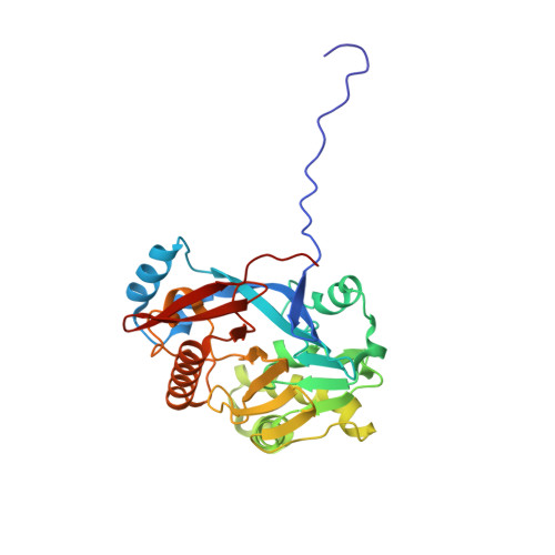
    6C2C

  • PubMed Abstract: 

    Characterizing the adaptive landscapes that encompass the emergence of novel enzyme functions can provide molecular insights into both enzymatic and evolutionary mechanisms. Here, we combine ancestral protein reconstruction with biochemical, structural and mutational analyses to characterize the functional evolution of methyl-parathion hydrolase (MPH), an organophosphate-degrading enzyme. We identify five mutations that are necessary and sufficient for the evolution of MPH from an ancestral dihydrocoumarin hydrolase. In-depth analyses of the adaptive landscapes encompassing this evolutionary transition revealed that the mutations form a complex interaction network, defined in part by higher-order epistasis, that constrained the adaptive pathways available. By also characterizing the adaptive landscapes in terms of their functional activities towards three additional organophosphate substrates, we reveal that subtle differences in the polarity of the substrate substituents drastically alter the network of epistatic interactions. Our work suggests that the mutations function collectively to enable substrate recognition via subtle structural repositioning.


  • Organizational Affiliation
    • Michael Smith Laboratories, University of British Columbia, Vancouver, British Columbia, Canada.

Macromolecule Content 

  • Total Structure Weight: 63.56 kDa 
  • Atom Count: 5,084 
  • Modeled Residue Count: 598 
  • Deposited Residue Count: 598 
  • Unique protein chains: 1

Macromolecules

Find similar proteins by:|  3D Structure
Entity ID: 1
MoleculeChains  Sequence LengthOrganismDetailsImage
dihydrocoumarin hydrolase, AncDHCH1
A, B
299Pseudomonas sp.Mutation(s): 0 

Small Molecules

Ligands 3 Unique
IDChains Name / Formula / InChI Key2D Diagram3D Interactions
PEG

Query on PEG


Download:Ideal Coordinates CCD File
F [auth A],
J [auth B]
DI(HYDROXYETHYL)ETHER
C4 H10 O3
MTHSVFCYNBDYFN-UHFFFAOYSA-N
ZN

Query on ZN


Download:Ideal Coordinates CCD File
C [auth A],
D [auth A],
G [auth B],
H [auth B]
ZINC ION
Zn
PTFCDOFLOPIGGS-UHFFFAOYSA-N
MG

Query on MG


Download:Ideal Coordinates CCD File
E [auth A],
I [auth B]
MAGNESIUM ION
Mg
JLVVSXFLKOJNIY-UHFFFAOYSA-N

Experimental Data & Validation

Experimental Data

  • Method: X-RAY DIFFRACTION
  • Resolution: 1.60 Å
  • R-Value Free:  0.204 (Depositor), 0.205 (DCC) 
  • R-Value Work:  0.173 (Depositor), 0.175 (DCC) 
  • R-Value Observed: 0.175 (Depositor) 
Space Group: P 21 21 21
Unit Cell:
Length ( Å )Angle ( ˚ )
a = 57.6α = 90
b = 88.15β = 90
c = 119.8γ = 90
Software Package:
Software NamePurpose
PHENIXrefinement
XDSdata reduction
Aimlessdata scaling
MOLREPphasing

Structure Validation

View Full Validation Report


Entry History 

Deposition Data

Revision History  (Full details and data files)

  • Version 1.0: 2019-01-16
    Type: Initial release
  • Version 1.1: 2019-01-23
    Changes: Data collection, Database references
  • Version 1.2: 2019-11-06
    Changes: Data collection, Database references
  • Version 1.3: 2024-03-13
    Changes: Data collection, Database references, Derived calculations