6BRA | pdb_00006bra

HIV-1 protease (D25N, inactive) in complex with phage display optimized substrate SGIFLETS


Experimental Data Snapshot

  • Method: X-RAY DIFFRACTION
  • Resolution: 1.11 Å
  • R-Value Free: 
    0.184 (Depositor), 0.185 (DCC) 
  • R-Value Work: 
    0.171 (Depositor), 0.171 (DCC) 
  • R-Value Observed: 
    0.171 (Depositor) 

Starting Model: experimental
View more details

wwPDB Validation 3D Report Full Report

Validation slider image for 6BRA

This is version 1.3 of the entry. See complete history

Literature

A substrate selected by phage display exhibits enhanced side-chain hydrogen bonding to HIV-1 protease.

Windsor, I.W.Raines, R.T.

(2018) Acta Crystallogr D Struct Biol 74: 690-694

  • DOI: https://doi.org/10.1107/S2059798318006691
  • Primary Citation Related Structures: 
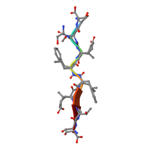
    6BRA

  • PubMed Abstract: 

    Crystal structures of inactive variants of HIV-1 protease bound to peptides have revealed how the enzyme recognizes its endogenous substrates. The best of the known substrates is, however, a nonnatural substrate that was identified by directed evolution. The crystal structure of the complex between this substrate and the D25N variant of the protease is reported at a resolution of 1.1 Å. The structure has several unprecedented features, especially the formation of additional hydrogen bonds between the enzyme and the substrate. This work expands the understanding of molecular recognition by HIV-1 protease and informs the design of new substrates and inhibitors.


  • Organizational Affiliation
    • Department of Chemistry, Massachusetts Institute of Technology, Cambridge, MA 02139, USA.

Macromolecule Content 

  • Total Structure Weight: 22.47 kDa 
  • Atom Count: 2,101 
  • Modeled Residue Count: 206 
  • Deposited Residue Count: 206 
  • Unique protein chains: 2

Macromolecules

Find similar proteins by:|  3D Structure
Entity ID: 1
MoleculeChains  Sequence LengthOrganismDetailsImage
Protease
A, B
99Human immunodeficiency virus 1Mutation(s): 6 
Gene Names: pol
UniProt
Find proteins for Q5RZ08 (Human immunodeficiency virus type 1)
Explore Q5RZ08 
Go to UniProtKB:  Q5RZ08
Entity Groups
Sequence Clusters30% Identity50% Identity70% Identity90% Identity95% Identity100% Identity
UniProt GroupQ5RZ08
Sequence Annotations
Expand
Reference Sequence
Find similar proteins by:  Sequence   |   3D Structure  
Entity ID: 2
MoleculeChains  Sequence LengthOrganismDetailsImage
Phage display-optimized HIV-1 protease substrateC [auth S]8Enterobacteria phage fdMutation(s): 0 

Experimental Data & Validation

Experimental Data

  • Method: X-RAY DIFFRACTION
  • Resolution: 1.11 Å
  • R-Value Free:  0.184 (Depositor), 0.185 (DCC) 
  • R-Value Work:  0.171 (Depositor), 0.171 (DCC) 
  • R-Value Observed: 0.171 (Depositor) 
Space Group: P 21 21 2
Unit Cell:
Length ( Å )Angle ( ˚ )
a = 58.033α = 90
b = 85.767β = 90
c = 46.13γ = 90
Software Package:
Software NamePurpose
HKL-2000data reduction
PHENIXrefinement
PDB_EXTRACTdata extraction
SCALEPACKdata scaling
PHASERphasing

Structure Validation

View Full Validation Report


Entry History 

& Funding Information

Deposition Data


Funding OrganizationLocationGrant Number
National Institutes of Health/National Institute of General Medical Sciences (NIH/NIGMS)United StatesP41 GM103399
National Institutes of Health/National Institute of General Medical Sciences (NIH/NIGMS)United StatesR01 GM044783

Revision History  (Full details and data files)

  • Version 1.0: 2018-07-18
    Type: Initial release
  • Version 1.1: 2019-02-20
    Changes: Author supporting evidence, Data collection
  • Version 1.2: 2020-01-01
    Changes: Author supporting evidence
  • Version 1.3: 2023-10-04
    Changes: Data collection, Database references, Refinement description