6BGD

The crystal structure of the W145A variant of TpMglB-2 (Tp0684) with bound ligand


Experimental Data Snapshot

  • Method: X-RAY DIFFRACTION
  • Resolution: 1.47 Å
  • R-Value Free: 0.144 
  • R-Value Work: 0.114 
  • R-Value Observed: 0.115 

wwPDB Validation   3D Report Full Report


This is version 1.6 of the entry. See complete history


Literature

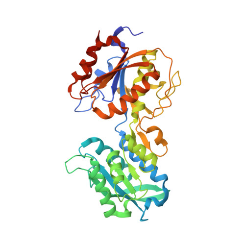
Crystal structures of MglB-2 (TP0684), a topologically variant d-glucose-binding protein from Treponema pallidum, reveal a ligand-induced conformational change.

Brautigam, C.A.Deka, R.K.Liu, W.Z.Norgard, M.V.

(2018) Protein Sci 27: 880-885

  • DOI: https://doi.org/10.1002/pro.3373
  • Primary Citation of Related Structures:  
    6BGC, 6BGD

  • PubMed Abstract: 

    Previously, we determined the crystal structure of apo-TpMglB-2, a d-glucose-binding component of a putative ABC transporter from the syphilis spirochete Treponema pallidum. The protein had an unusual topology for this class of proteins, raising the question of whether the d-glucose-binding mode would be different in TpMglB-2. Here, we present the crystal structures of a variant of TpMglB-2 with and without d-glucose bound. The structures demonstrate that, despite its aberrant topology, the protein undergoes conformational changes and binds d-glucose similarly to other Mgl-type proteins, likely facilitating d-glucose uptake in T. pallidum.


  • Organizational Affiliation

    Department of Biophysics, The University of Texas Southwestern Medical Center, Dallas, Texas, 75390.


Macromolecules
Find similar proteins by:  (by identity cutoff)  |  3D Structure
Entity ID: 1
MoleculeChains Sequence LengthOrganismDetailsImage
Glucose/galactose-binding lipoprotein378Treponema pallidum subsp. pallidum str. NicholsMutation(s): 1 
Gene Names: mglBtpp38TP_0684
UniProt
Find proteins for Q08255 (Treponema pallidum (strain Nichols))
Explore Q08255 
Go to UniProtKB:  Q08255
Entity Groups  
Sequence Clusters30% Identity50% Identity70% Identity90% Identity95% Identity100% Identity
UniProt GroupQ08255
Sequence Annotations
Expand
  • Reference Sequence
Small Molecules
Ligands 2 Unique
IDChains Name / Formula / InChI Key2D Diagram3D Interactions
EDO
Query on EDO

Download Ideal Coordinates CCD File 
B [auth A]
C [auth A]
D [auth A]
E [auth A]
F [auth A]
B [auth A],
C [auth A],
D [auth A],
E [auth A],
F [auth A],
G [auth A],
H [auth A],
I [auth A],
J [auth A],
K [auth A]
1,2-ETHANEDIOL
C2 H6 O2
LYCAIKOWRPUZTN-UHFFFAOYSA-N
CA
Query on CA

Download Ideal Coordinates CCD File 
L [auth A]CALCIUM ION
Ca
BHPQYMZQTOCNFJ-UHFFFAOYSA-N
Experimental Data & Validation

Experimental Data

  • Method: X-RAY DIFFRACTION
  • Resolution: 1.47 Å
  • R-Value Free: 0.144 
  • R-Value Work: 0.114 
  • R-Value Observed: 0.115 
  • Space Group: C 2 2 21
Unit Cell:
Length ( Å )Angle ( ˚ )
a = 75.834α = 90
b = 110.199β = 90
c = 89.304γ = 90
Software Package:
Software NamePurpose
PHENIXrefinement
HKL-3000data reduction
HKL-3000data scaling
PHASERphasing

Structure Validation

View Full Validation Report


Entry History & Funding Information

Deposition Data


Funding OrganizationLocationGrant Number
National Institutes of Health/National Institute Of Allergy and Infectious Diseases (NIH/NIAID)United StatesAI056305

Revision History  (Full details and data files)

  • Version 1.0: 2018-01-31
    Type: Initial release
  • Version 1.1: 2018-02-14
    Changes: Author supporting evidence
  • Version 1.2: 2018-04-04
    Changes: Data collection, Database references
  • Version 1.3: 2019-08-21
    Changes: Data collection, Database references
  • Version 1.4: 2019-12-11
    Changes: Author supporting evidence
  • Version 1.5: 2023-02-15
    Changes: Database references, Structure summary
  • Version 1.6: 2023-10-25
    Changes: Data collection, Refinement description