6B4J

Crystal structure of human Gle1 CTD-Nup42 GBM-DDX19B(AMPPNP) complex


Experimental Data Snapshot

  • Method: X-RAY DIFFRACTION
  • Resolution: 3.40 Å
  • R-Value Free: 0.313 
  • R-Value Work: 0.261 
  • R-Value Observed: 0.266 

wwPDB Validation   3D Report Full Report


Ligand Structure Quality Assessment 


This is version 1.3 of the entry. See complete history


Literature

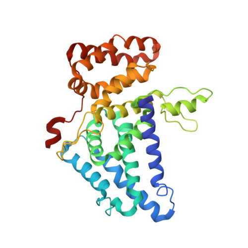
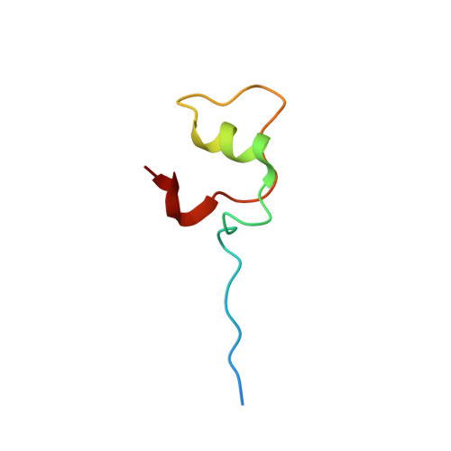
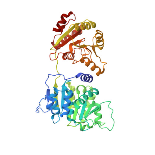
Structural and functional analysis of mRNA export regulation by the nuclear pore complex.

Lin, D.H.Correia, A.R.Cai, S.W.Huber, F.M.Jette, C.A.Hoelz, A.

(2018) Nat Commun 9: 2319-2319

  • DOI: https://doi.org/10.1038/s41467-018-04459-3
  • Primary Citation of Related Structures:  
    6B4E, 6B4F, 6B4G, 6B4H, 6B4I, 6B4J, 6B4K

  • PubMed Abstract: 

    The nuclear pore complex (NPC) controls the passage of macromolecules between the nucleus and cytoplasm, but how the NPC directly participates in macromolecular transport remains poorly understood. In the final step of mRNA export, the DEAD-box helicase DDX19 is activated by the nucleoporins Gle1, Nup214, and Nup42 to remove Nxf1•Nxt1 from mRNAs. Here, we report crystal structures of Gle1•Nup42 from three organisms that reveal an evolutionarily conserved binding mode. Biochemical reconstitution of the DDX19 ATPase cycle establishes that human DDX19 activation does not require IP 6 , unlike its fungal homologs, and that Gle1 stability affects DDX19 activation. Mutations linked to motor neuron diseases cause decreased Gle1 thermostability, implicating nucleoporin misfolding as a disease determinant. Crystal structures of human Gle1•Nup42•DDX19 reveal the structural rearrangements in DDX19 from an auto-inhibited to an RNA-binding competent state. Together, our results provide the foundation for further mechanistic analyses of mRNA export in humans.


  • Organizational Affiliation

    Division of Chemistry and Chemical Engineering, California Institute of Technology, 1200 East California Boulevard, Pasadena, CA, 91125, USA.


Macromolecules
Find similar proteins by:  (by identity cutoff)  |  3D Structure
Entity ID: 1
MoleculeChains Sequence LengthOrganismDetailsImage
Nucleoporin GLE1A [auth B],
C [auth A]
317Homo sapiensMutation(s): 0 
Gene Names: GLE1GLE1L
UniProt & NIH Common Fund Data Resources
Find proteins for Q53GS7 (Homo sapiens)
Explore Q53GS7 
Go to UniProtKB:  Q53GS7
PHAROS:  Q53GS7
GTEx:  ENSG00000119392 
Entity Groups  
Sequence Clusters30% Identity50% Identity70% Identity90% Identity95% Identity100% Identity
UniProt GroupQ53GS7
Sequence Annotations
Expand
  • Reference Sequence
Find similar proteins by:  (by identity cutoff)  |  3D Structure
Entity ID: 2
MoleculeChains Sequence LengthOrganismDetailsImage
Nucleoporin like 2B [auth C],
D
49Homo sapiensMutation(s): 0 
Gene Names: NUPL2
UniProt & NIH Common Fund Data Resources
Find proteins for O15504 (Homo sapiens)
Explore O15504 
Go to UniProtKB:  O15504
PHAROS:  O15504
GTEx:  ENSG00000136243 
Entity Groups  
Sequence Clusters30% Identity50% Identity70% Identity90% Identity95% Identity100% Identity
UniProt GroupO15504
Sequence Annotations
Expand
  • Reference Sequence
Find similar proteins by:  (by identity cutoff)  |  3D Structure
Entity ID: 3
MoleculeChains Sequence LengthOrganismDetailsImage
ATP-dependent RNA helicase DDX19B
E, F
430Homo sapiensMutation(s): 0 
Gene Names: DDX19BDBP5DDX19TDBP
EC: 3.6.4.13
UniProt & NIH Common Fund Data Resources
Find proteins for Q9UMR2 (Homo sapiens)
Explore Q9UMR2 
Go to UniProtKB:  Q9UMR2
PHAROS:  Q9UMR2
GTEx:  ENSG00000157349 
Entity Groups  
Sequence Clusters30% Identity50% Identity70% Identity90% Identity95% Identity100% Identity
UniProt GroupQ9UMR2
Sequence Annotations
Expand
  • Reference Sequence
Small Molecules
Ligands 3 Unique
IDChains Name / Formula / InChI Key2D Diagram3D Interactions
ANP
Query on ANP

Download Ideal Coordinates CCD File 
L [auth E],
P [auth F]
PHOSPHOAMINOPHOSPHONIC ACID-ADENYLATE ESTER
C10 H17 N6 O12 P3
PVKSNHVPLWYQGJ-KQYNXXCUSA-N
PO4
Query on PO4

Download Ideal Coordinates CCD File 
G [auth B]
H [auth B]
I [auth E]
J [auth E]
M [auth F]
G [auth B],
H [auth B],
I [auth E],
J [auth E],
M [auth F],
N [auth F]
PHOSPHATE ION
O4 P
NBIIXXVUZAFLBC-UHFFFAOYSA-K
MG
Query on MG

Download Ideal Coordinates CCD File 
K [auth E],
O [auth F]
MAGNESIUM ION
Mg
JLVVSXFLKOJNIY-UHFFFAOYSA-N
Experimental Data & Validation

Experimental Data

  • Method: X-RAY DIFFRACTION
  • Resolution: 3.40 Å
  • R-Value Free: 0.313 
  • R-Value Work: 0.261 
  • R-Value Observed: 0.266 
  • Space Group: P 1 21 1
Unit Cell:
Length ( Å )Angle ( ˚ )
a = 87.94α = 90
b = 73.56β = 95.07
c = 145.86γ = 90
Software Package:
Software NamePurpose
PHENIXrefinement
DIALSdata reduction
Aimlessdata scaling
PHASERphasing

Structure Validation

View Full Validation Report


Ligand Structure Quality Assessment 


Entry History 

Deposition Data

Revision History  (Full details and data files)

  • Version 1.0: 2018-06-20
    Type: Initial release
  • Version 1.1: 2018-06-27
    Changes: Data collection, Database references
  • Version 1.2: 2018-11-21
    Changes: Data collection
  • Version 1.3: 2024-03-13
    Changes: Data collection, Database references, Derived calculations