6AYY

Crystal structure of Apo fructose-1,6-bisphosphatase from Mycobacterium tuberculosis


Experimental Data Snapshot

  • Method: X-RAY DIFFRACTION
  • Resolution: 2.60 Å
  • R-Value Free: 0.259 
  • R-Value Work: 0.191 
  • R-Value Observed: 0.194 

wwPDB Validation   3D Report Full Report


This is version 1.3 of the entry. See complete history


Literature

Structures of the Mycobacterium tuberculosis GlpX protein (class II fructose-1,6-bisphosphatase): implications for the active oligomeric state, catalytic mechanism and citrate inhibition.

Wolf, N.M.Gutka, H.J.Movahedzadeh, F.Abad-Zapatero, C.

(2018) Acta Crystallogr D Struct Biol 74: 321-331

  • DOI: https://doi.org/10.1107/S2059798318002838
  • Primary Citation of Related Structures:  
    6AYY

  • PubMed Abstract: 

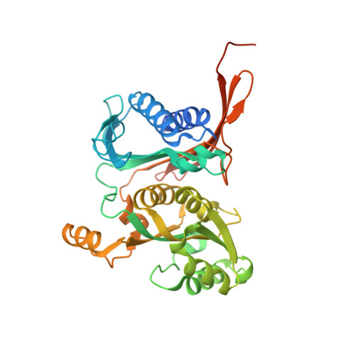
    The crystal structures of native class II fructose-1,6-bisphosphatase (FBPaseII) from Mycobacterium tuberculosis at 2.6 Å resolution and two active-site protein variants are presented. The variants were complexed with the reaction product fructose 6-phosphate (F6P). The Thr84Ala mutant is inactive, while the Thr84Ser mutant has a lower catalytic activity. The structures reveal the presence of a 222 tetramer, similar to those described for fructose-1,6/sedoheptulose-1,7-bisphosphatase from Synechocystis (strain 6803) as well as the equivalent enzyme from Thermosynechococcus elongatus. This homotetramer corresponds to a homologous oligomer that is present but not described in the crystal structure of FBPaseII from Escherichia coli and is probably conserved in all FBPaseIIs. The constellation of amino-acid residues in the active site of FBPaseII from M. tuberculosis (MtFBPaseII) is conserved and is analogous to that described previously for the E. coli enzyme. Moreover, the structure of the active site of the partially active (Thr84Ser) variant and the analysis of the kinetics are consistent with the previously proposed catalytic mechanism. The presence of metabolites in the crystallization medium (for example citrate and malonate) and in the corresponding crystal structures of MtFBPaseII, combined with their observed inhibitory effect, could suggest the existence of an uncharacterized inhibition of this class of enzymes besides the allosteric inhibition by adenosine monophosphate observed for the Synechocystis enzyme. The structural and functional insights derived from the structure of MtFBPaseII will provide critical information for the design of lead inhibitors, which will be used to validate this target for future chemical intervention.


  • Organizational Affiliation

    Institute for Tuberculosis Research, University of Illinois at Chicago, Chicago, Illinois, USA.


Macromolecules
Find similar proteins by:  (by identity cutoff)  |  3D Structure
Entity ID: 1
MoleculeChains Sequence LengthOrganismDetailsImage
Fructose-1,6-bisphosphatase class 2
A, B
347Mycobacterium tuberculosis CDC1551Mutation(s): 0 
Gene Names: glpXMT1131
EC: 3.1.3.11
UniProt
Find proteins for P9WN21 (Mycobacterium tuberculosis (strain ATCC 25618 / H37Rv))
Explore P9WN21 
Go to UniProtKB:  P9WN21
Entity Groups  
Sequence Clusters30% Identity50% Identity70% Identity90% Identity95% Identity100% Identity
UniProt GroupP9WN21
Sequence Annotations
Expand
  • Reference Sequence
Small Molecules
Ligands 3 Unique
IDChains Name / Formula / InChI Key2D Diagram3D Interactions
CIT
Query on CIT

Download Ideal Coordinates CCD File 
W [auth B]CITRIC ACID
C6 H8 O7
KRKNYBCHXYNGOX-UHFFFAOYSA-N
GOL
Query on GOL

Download Ideal Coordinates CCD File 
C [auth A]
D [auth A]
E [auth A]
F [auth A]
G [auth A]
C [auth A],
D [auth A],
E [auth A],
F [auth A],
G [auth A],
H [auth A],
I [auth A],
K [auth A],
L [auth A],
M [auth B],
N [auth B],
O [auth B],
P [auth B],
Q [auth B],
R [auth B],
S [auth B],
T [auth B],
U [auth B],
V [auth B],
X [auth B]
GLYCEROL
C3 H8 O3
PEDCQBHIVMGVHV-UHFFFAOYSA-N
UNX
Query on UNX

Download Ideal Coordinates CCD File 
J [auth A]UNKNOWN ATOM OR ION
X
Experimental Data & Validation

Experimental Data

  • Method: X-RAY DIFFRACTION
  • Resolution: 2.60 Å
  • R-Value Free: 0.259 
  • R-Value Work: 0.191 
  • R-Value Observed: 0.194 
  • Space Group: P 61 2 2
Unit Cell:
Length ( Å )Angle ( ˚ )
a = 131.541α = 90
b = 131.541β = 90
c = 144.352γ = 120
Software Package:
Software NamePurpose
PHENIXrefinement
HKL-2000data reduction
HKL-2000data scaling
MOLREPphasing

Structure Validation

View Full Validation Report


Entry History & Funding Information

Deposition Data


Funding OrganizationLocationGrant Number
Potts Memorial FoundationUnited StatesG3541

Revision History  (Full details and data files)

  • Version 1.0: 2018-03-14
    Type: Initial release
  • Version 1.1: 2018-04-25
    Changes: Data collection, Database references
  • Version 1.2: 2019-03-27
    Changes: Data collection, Derived calculations
  • Version 1.3: 2023-10-04
    Changes: Data collection, Database references, Refinement description