6AU1

Structure of the PgaB (BpsB) glycoside hydrolase domain from Bordetella bronchiseptica


Experimental Data Snapshot

  • Method: X-RAY DIFFRACTION
  • Resolution: 1.76 Å
  • R-Value Free: 0.167 
  • R-Value Work: 0.151 
  • R-Value Observed: 0.151 

wwPDB Validation   3D Report Full Report


This is version 1.3 of the entry. See complete history


Literature

PgaB orthologues contain a glycoside hydrolase domain that cleaves deacetylated poly-beta (1,6)-N-acetylglucosamine and can disrupt bacterial biofilms.

Little, D.J.Pfoh, R.Le Mauff, F.Bamford, N.C.Notte, C.Baker, P.Guragain, M.Robinson, H.Pier, G.B.Nitz, M.Deora, R.Sheppard, D.C.Howell, P.L.

(2018) PLoS Pathog 14: e1006998-e1006998

  • DOI: https://doi.org/10.1371/journal.ppat.1006998
  • Primary Citation of Related Structures:  
    6AU1

  • PubMed Abstract: 

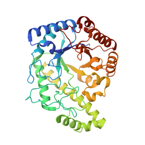
    Poly-β(1,6)-N-acetyl-D-glucosamine (PNAG) is a major biofilm component of many pathogenic bacteria. The production, modification, and export of PNAG in Escherichia coli and Bordetella species require the protein products encoded by the pgaABCD operon. PgaB is a two-domain periplasmic protein that contains an N-terminal deacetylase domain and a C-terminal PNAG binding domain that is critical for export. However, the exact function of the PgaB C-terminal domain remains unclear. Herein, we show that the C-terminal domains of Bordetella bronchiseptica PgaB (PgaBBb) and E. coli PgaB (PgaBEc) function as glycoside hydrolases. These enzymes hydrolyze purified deacetylated PNAG (dPNAG) from Staphylococcus aureus, disrupt PNAG-dependent biofilms formed by Bordetella pertussis, Staphylococcus carnosus, Staphylococcus epidermidis, and E. coli, and potentiate bacterial killing by gentamicin. Furthermore, we found that PgaBBb was only able to hydrolyze PNAG produced in situ by the E. coli PgaCD synthase complex when an active deacetylase domain was present. Mass spectrometry analysis of the PgaB-hydrolyzed dPNAG substrate showed a GlcN-GlcNAc-GlcNAc motif at the new reducing end of detected fragments. Our 1.76 Å structure of the C-terminal domain of PgaBBb reveals a central cavity within an elongated surface groove that appears ideally suited to recognize the GlcN-GlcNAc-GlcNAc motif. The structure, in conjunction with molecular modeling and site directed mutagenesis led to the identification of the dPNAG binding subsites and D474 as the probable catalytic acid. This work expands the role of PgaB within the PNAG biosynthesis machinery, defines a new glycoside hydrolase family GH153, and identifies PgaB as a possible therapeutic agent for treating PNAG-dependent biofilm infections.


  • Organizational Affiliation

    Program in Molecular Medicine, The Hospital for Sick Children, Toronto, ON, Canada.


Macromolecules
Find similar proteins by:  (by identity cutoff)  |  3D Structure
Entity ID: 1
MoleculeChains Sequence LengthOrganismDetailsImage
Putative hemin storage protein
A, B
357Bordetella bronchiseptica RB50Mutation(s): 0 
Gene Names: hmsFBB1768PgaB (BpsBHmsF)
EC: 3.2.1
UniProt
Find proteins for A0A0H3LKK6 (Bordetella bronchiseptica (strain ATCC BAA-588 / NCTC 13252 / RB50))
Explore A0A0H3LKK6 
Go to UniProtKB:  A0A0H3LKK6
Entity Groups  
Sequence Clusters30% Identity50% Identity70% Identity90% Identity95% Identity100% Identity
UniProt GroupA0A0H3LKK6
Sequence Annotations
Expand
  • Reference Sequence
Small Molecules
Ligands 3 Unique
IDChains Name / Formula / InChI Key2D Diagram3D Interactions
SO4
Query on SO4

Download Ideal Coordinates CCD File 
C [auth A]
D [auth A]
E [auth A]
F [auth A]
H [auth B]
C [auth A],
D [auth A],
E [auth A],
F [auth A],
H [auth B],
I [auth B],
J [auth B],
K [auth B]
SULFATE ION
O4 S
QAOWNCQODCNURD-UHFFFAOYSA-L
EDO
Query on EDO

Download Ideal Coordinates CCD File 
G [auth A],
M [auth B],
N [auth B],
O [auth B]
1,2-ETHANEDIOL
C2 H6 O2
LYCAIKOWRPUZTN-UHFFFAOYSA-N
CL
Query on CL

Download Ideal Coordinates CCD File 
L [auth B]CHLORIDE ION
Cl
VEXZGXHMUGYJMC-UHFFFAOYSA-M
Experimental Data & Validation

Experimental Data

  • Method: X-RAY DIFFRACTION
  • Resolution: 1.76 Å
  • R-Value Free: 0.167 
  • R-Value Work: 0.151 
  • R-Value Observed: 0.151 
  • Space Group: P 1 21 1
Unit Cell:
Length ( Å )Angle ( ˚ )
a = 72.102α = 90
b = 91.649β = 111.51
c = 75.295γ = 90
Software Package:
Software NamePurpose
PHENIXrefinement
HKL-2000data reduction
SCALEPACKdata scaling
PHENIXphasing

Structure Validation

View Full Validation Report


Entry History & Funding Information

Deposition Data


Funding OrganizationLocationGrant Number
Canadian Institutes of Health Research (CIHR)Canada43998

Revision History  (Full details and data files)

  • Version 1.0: 2018-04-18
    Type: Initial release
  • Version 1.1: 2018-05-16
    Changes: Data collection, Database references
  • Version 1.2: 2020-01-08
    Changes: Author supporting evidence
  • Version 1.3: 2023-10-04
    Changes: Data collection, Database references, Refinement description