6AQ7

Structure of POM6 FAB fragment complexed with mouse PrPc


Experimental Data Snapshot

  • Method: X-RAY DIFFRACTION
  • Resolution: 1.83 Å
  • R-Value Free: 0.230 
  • R-Value Work: 0.197 
  • R-Value Observed: 0.198 

wwPDB Validation   3D Report Full Report


Ligand Structure Quality Assessment 


This is version 1.3 of the entry. See complete history


Literature

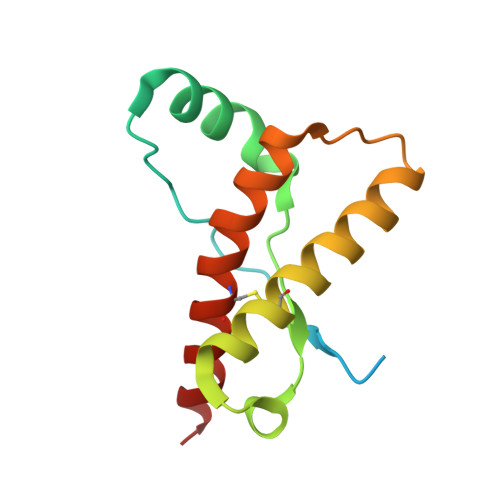
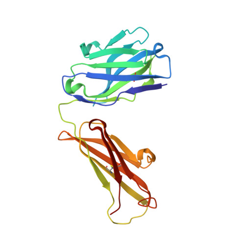
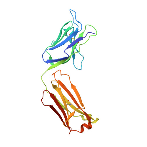
Structural characterization of POM6 Fab and mouse prion protein complex identifies key regions for prions conformational conversion.

Baral, P.K.Swayampakula, M.Aguzzi, A.James, M.N.G.

(2018) FEBS J 285: 1701-1714

  • DOI: https://doi.org/10.1111/febs.14438
  • Primary Citation of Related Structures:  
    6AQ7

  • PubMed Abstract: 

    Conversion of the cellular prion protein PrP C into its pathogenic isoform PrP S c is the hallmark of prion diseases, fatal neurodegenerative diseases affecting many mammalian species including humans. Anti-prion monoclonal antibodies can arrest the progression of prion diseases by stabilizing the cellular form of the prion protein. Here, we present the crystal structure of the POM6 Fab fragment, in complex with the mouse prion protein (moPrP). The prion epitope of POM6 is in close proximity to the epitope recognized by the purportedly toxic antibody fragment, POM1 Fab also complexed with moPrP. The POM6 Fab recognizes a larger binding interface indicating a likely stronger binding compared to POM1. POM6 and POM1 exhibit distinct biological responses. Structural comparisons of the bound mouse prion proteins from the POM6 Fab:moPrP and POM1 Fab:moPrP complexes reveal several key regions of the prion protein that might be involved in initiating mis-folding events.


  • Organizational Affiliation

    Department of Biochemistry, Faculty of Medicine and Dentistry, University of Alberta, Edmonton, Canada.


Macromolecules
Find similar proteins by:  (by identity cutoff)  |  3D Structure
Entity ID: 1
MoleculeChains Sequence LengthOrganismDetailsImage
Major prion protein122Mus musculusMutation(s): 0 
Gene Names: PrnpPrn-pPrp
UniProt
Find proteins for P04925 (Mus musculus)
Explore P04925 
Go to UniProtKB:  P04925
Entity Groups  
Sequence Clusters30% Identity50% Identity70% Identity90% Identity95% Identity100% Identity
UniProt GroupP04925
Sequence Annotations
Expand
  • Reference Sequence
Find similar proteins by:  (by identity cutoff)  |  3D Structure
Entity ID: 2
MoleculeChains Sequence LengthOrganismDetailsImage
POM6 FAB Heavy CHAINB [auth H]214Mus musculusMutation(s): 0 
Entity Groups  
Sequence Clusters30% Identity50% Identity70% Identity90% Identity95% Identity100% Identity
Sequence Annotations
Expand
  • Reference Sequence
Find similar proteins by:  (by identity cutoff)  |  3D Structure
Entity ID: 3
MoleculeChains Sequence LengthOrganismDetailsImage
POM6 FAB light CHAINC [auth L]216Mus musculusMutation(s): 0 
Entity Groups  
Sequence Clusters30% Identity50% Identity70% Identity90% Identity95% Identity100% Identity
Sequence Annotations
Expand
  • Reference Sequence
Experimental Data & Validation

Experimental Data

  • Method: X-RAY DIFFRACTION
  • Resolution: 1.83 Å
  • R-Value Free: 0.230 
  • R-Value Work: 0.197 
  • R-Value Observed: 0.198 
  • Space Group: P 2 21 21
Unit Cell:
Length ( Å )Angle ( ˚ )
a = 72.04α = 90
b = 82.42β = 90
c = 103.7γ = 90
Software Package:
Software NamePurpose
PHENIXrefinement
XDSdata reduction
XDSdata scaling
MOLREPphasing

Structure Validation

View Full Validation Report


Ligand Structure Quality Assessment 


Entry History & Funding Information

Deposition Data


Funding OrganizationLocationGrant Number
Alberta innovatesCanada--

Revision History  (Full details and data files)

  • Version 1.0: 2018-04-04
    Type: Initial release
  • Version 1.1: 2018-05-16
    Changes: Data collection, Database references
  • Version 1.2: 2018-11-07
    Changes: Data collection, Source and taxonomy
  • Version 1.3: 2023-10-04
    Changes: Data collection, Database references, Refinement description