6APZ | pdb_00006apz

Crystal structure of Mycobacterium tuberculosis malate synthase in complex with 3-hydroxy-phenyldiketoacid


Experimental Data Snapshot

  • Method: X-RAY DIFFRACTION
  • Resolution: 2.25 Å
  • R-Value Free: 
    0.255 (Depositor), 0.253 (DCC) 
  • R-Value Work: 
    0.178 (Depositor), 0.179 (DCC) 
  • R-Value Observed: 
    0.182 (Depositor) 

Starting Model: experimental
View more details

wwPDB Validation 3D Report Full Report

Validation slider image for 6APZ

Ligand Structure Quality Assessment 


This is version 1.4 of the entry. See complete history

Literature

Anion-pi Interactions in Computer-Aided Drug Design: Modeling the Inhibition of Malate Synthase by Phenyl-Diketo Acids.

Ellenbarger, J.F.Krieger, I.V.Huang, H.L.Gomez-Coca, S.Ioerger, T.R.Sacchettini, J.C.Wheeler, S.E.Dunbar, K.R.

(2018) J Chem Inf Model 58: 2085-2091

  • DOI: https://doi.org/10.1021/acs.jcim.8b00417
  • Primary Citation Related Structures: 
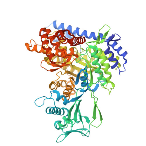
    6APZ, 6AS6, 6ASU, 6AU9, 6AXB, 6BA7, 6BU1, 6C2X, 6C6O, 6C7B, 6C8P, 6DKO, 6DL9, 6DLJ, 6DNP

  • PubMed Abstract: 

    Human infection by Mycobacterium tuberculosis (Mtb) continues to be a global epidemic. Computer-aided drug design (CADD) methods are used to accelerate traditional drug discovery efforts. One noncovalent interaction that is being increasingly identified in biological systems but is neglected in CADD is the anion-π interaction. The study reported herein supports the conclusion that anion-π interactions play a central role in directing the binding of phenyl-diketo acid (PDKA) inhibitors to malate synthase (GlcB), an enzyme required for Mycobacterium tuberculosis virulence. Using density functional theory methods (M06-2X/6-31+G(d)), a GlcB active site template was developed for a predictive model through a comparative analysis of PDKA-bound GlcB crystal structures. The active site model includes the PDKA molecule and the protein determinants of the electrostatic, hydrogen-bonding, and anion-π interactions involved in binding. The predictive model accurately determines the Asp 633-PDKA structural position upon binding and precisely predicts the relative binding enthalpies of a series of 2-ortho halide-PDKAs to GlcB. A screening model was also developed to efficiently assess the propensity of each PDKA analog to participate in an anion-π interaction; this method is in good agreement with both the predictive model and the experimental binding enthalpies for the 2-ortho halide-PDKAs. With the screening and predictive models in hand, we have developed an efficient method for computationally screening and evaluating the binding enthalpy of variously substituted PDKA molecules. This study serves to illustrate the contribution of this overlooked interaction to binding affinity and demonstrates the importance of integrating anion-π interactions into structure-based CADD.


  • Organizational Affiliation
    • Department of Chemistry , Texas A&M University , P.O. Box 30012, College Station , Texas 77842 , United States.

Macromolecule Content 

  • Total Structure Weight: 81.17 kDa 
  • Atom Count: 5,786 
  • Modeled Residue Count: 707 
  • Deposited Residue Count: 741 
  • Unique protein chains: 1

Macromolecules

Find similar proteins by:|  3D Structure
Entity ID: 1
MoleculeChains  Sequence LengthOrganismDetailsImage
Malate synthase G741Mycobacterium tuberculosis H37RaMutation(s): 1 
Gene Names: glcBMRA_1848
EC: 2.3.3.9
UniProt
Find proteins for P9WK17 (Mycobacterium tuberculosis (strain ATCC 25618 / H37Rv))
Explore P9WK17 
Go to UniProtKB:  P9WK17
Entity Groups
Sequence Clusters30% Identity50% Identity70% Identity90% Identity95% Identity100% Identity
UniProt GroupP9WK17
Sequence Annotations
Expand
Reference Sequence

Small Molecules

Ligands 4 Unique
IDChains Name / Formula / InChI Key2D Diagram3D Interactions
BQ4
(Subject of Investigation/LOI)

Query on BQ4


Download:Ideal Coordinates CCD File
C [auth A](2Z)-2-hydroxy-4-(3-hydroxyphenyl)-4-oxobut-2-enoic acid
C10 H8 O5
WWHSBYDKAHIVPP-UITAMQMPSA-N
PEG

Query on PEG


Download:Ideal Coordinates CCD File
E [auth A],
F [auth A],
G [auth A],
H [auth A]
DI(HYDROXYETHYL)ETHER
C4 H10 O3
MTHSVFCYNBDYFN-UHFFFAOYSA-N
NI

Query on NI


Download:Ideal Coordinates CCD File
D [auth A]NICKEL (II) ION
Ni
VEQPNABPJHWNSG-UHFFFAOYSA-N
MG

Query on MG


Download:Ideal Coordinates CCD File
B [auth A]MAGNESIUM ION
Mg
JLVVSXFLKOJNIY-UHFFFAOYSA-N

Experimental Data & Validation

Experimental Data

  • Method: X-RAY DIFFRACTION
  • Resolution: 2.25 Å
  • R-Value Free:  0.255 (Depositor), 0.253 (DCC) 
  • R-Value Work:  0.178 (Depositor), 0.179 (DCC) 
  • R-Value Observed: 0.182 (Depositor) 
Space Group: P 43 21 2
Unit Cell:
Length ( Å )Angle ( ˚ )
a = 79.378α = 90
b = 79.378β = 90
c = 226.174γ = 90
Software Package:
Software NamePurpose
PHENIXrefinement
HKL-2000data reduction
HKL-2000data scaling
MOLREPphasing

Structure Validation

View Full Validation Report


Ligand Structure Quality Assessment 


Entry History 

& Funding Information

Deposition Data


Funding OrganizationLocationGrant Number
National Institutes of Health/National Institute Of Allergy and Infectious Diseases (NIH/NIAID)United StatesP01 AI095208

Revision History  (Full details and data files)

  • Version 1.0: 2018-08-22
    Type: Initial release
  • Version 1.1: 2018-09-05
    Changes: Data collection, Database references
  • Version 1.2: 2018-10-31
    Changes: Data collection, Database references
  • Version 1.3: 2022-03-23
    Changes: Author supporting evidence, Database references, Derived calculations
  • Version 1.4: 2023-10-04
    Changes: Data collection, Refinement description