Identification and characterization of Helicobacter pylori O-acetylserine-dependent cystathionine beta-synthase, a distinct member of the PLP-II family.
Devi, S., Tarique, K.F., Ali, M.F., Abdul Rehman, S.A., Gourinath, S.(2019) Mol Microbiol 112: 718-739
- PubMed: 31132312 Search on PubMed
- DOI: https://doi.org/10.1111/mmi.14315
- Primary Citation Related Structures:
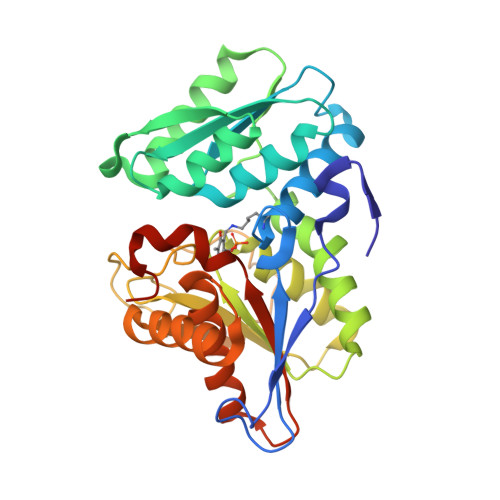
6AHI - PubMed Abstract:
O-acetylserine sulfhydrylase (OASS) and cystathionine β-synthase (CBS) are members of the PLP-II family, and involved in L-cysteine production. OASS produces L-cysteine via a de novo pathway while CBS participates in the reverse transsulfuration pathway. O-acetylserine-dependent CBS (OCBS) was previously identified as a new member of the PLP-II family, which are predominantly seen in bacteria. The bacterium Helicobacter pylori possess only one OASS (hp0107) gene and we showed that the protein coded by this gene actually functions as an OCBS and utilizes L-homocysteine and O-acetylserine (OAS) to produce cystathionine. HpOCBS did not show CBS activity with the substrate L-serine and required OAS exclusively. The HpOCBS structure in complex with methionine showed a closed cleft state, explaining the initial mode of substrate binding. Sequence and structural analyses showed differences between the active sites of OCBS and CBS, and explain their different substrate preferences. We identified three hydrophobic residues near the active site of OCBS, corresponding to one serine and two tyrosine residues in CBSs. Mutational studies were performed on HpOCBS and Saccharomyces cerevisiae CBS. A ScCBS double mutant (Y158F/Y226V) did not display activity with L-serine, indicating indispensability of these polar residues for selecting substrate L-serine, however, did show activity with OAS.
- Structural Biology Laboratory, School of Life Sciences, Jawaharlal Nehru University, New Delhi, 110067, India.
Organizational Affiliation: