6A4J

Crystal structure of Thioredoxin reductase 2 from Staphylococcus aureus


Experimental Data Snapshot

  • Method: X-RAY DIFFRACTION
  • Resolution: 3.40 Å
  • R-Value Free: 0.267 
  • R-Value Work: 0.241 
  • R-Value Observed: 0.243 

wwPDB Validation   3D Report Full Report


Ligand Structure Quality Assessment 


This is version 1.3 of the entry. See complete history


Literature

Elucidation of the mechanism of disulfide exchange between staphylococcal thioredoxin2 and thioredoxin reductase2: A structural insight.

Bose, M.Bhattacharyya, S.Biswas, R.Roychowdhury, A.Bhattacharjee, A.Ghosh, A.K.Das, A.K.

(2019) Biochimie 160: 1-13

  • DOI: https://doi.org/10.1016/j.biochi.2019.01.019
  • Primary Citation of Related Structures:  
    4RUV, 6A4J

  • PubMed Abstract: 

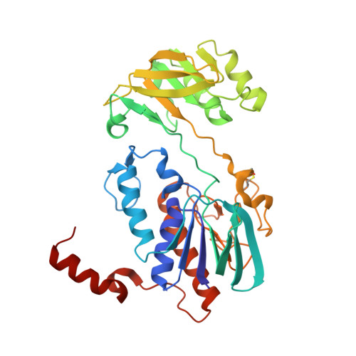
    The redox homeostasis of cytoplasm is maintained by a series of disulfide exchange reactions mediated by proteins belonging to the thioredoxin superfamily. Thioredoxin and thioredoxin reductase, being the major members of the family, play a key role in oxidative stress response of Staphylococcus aureus. In this report, we have identified and characterised an active thioredoxin system of the mentioned pathogen. Crystal structure of thioredoxin2 (SaTrx2) in its reduced form reveals that it contains the conserved redox active WCXXC motif and a thioredoxin fold. Thioredoxin reductase2 (SaTR2) is a flavoprotein and consists of two Rossmann folds as the binding sites for FAD and NADPH. Crystal structure of the SaTR2 holoenzyme shows that the protein consists of two domains and the catalytic site comprises of an intramolecular disulfide bond formed between two sequentially distal cysteine residues. Biophysical and biochemical studies unveil that SaTrx2 and SaTR2 can physically interact in solution and in the course of sustaining the redox equilibrium, the latter reduces the former. Molecular docking has been performed to illustrate the interface formed between SaTrx2 and SaTR2 during the disulfide exchange reaction.


  • Organizational Affiliation

    Department of Biotechnology, Indian Institute of Technology Kharagpur, Kharagpur, 721302, India.


Macromolecules
Find similar proteins by:  (by identity cutoff)  |  3D Structure
Entity ID: 1
MoleculeChains Sequence LengthOrganismDetailsImage
Ferredoxin--NADP reductase
A, B
344Staphylococcus aureusMutation(s): 0 
Gene Names: trxB_2EP54_04310EQ90_00075RK60_12235RK64_12650RK68_02995RK73_09520RK98_08250RL06_05030SAMEA3448991_00661
EC: 1.18.1.2
UniProt
Find proteins for Q2FVP8 (Staphylococcus aureus (strain NCTC 8325 / PS 47))
Explore Q2FVP8 
Go to UniProtKB:  Q2FVP8
Entity Groups  
Sequence Clusters30% Identity50% Identity70% Identity90% Identity95% Identity100% Identity
UniProt GroupQ2FVP8
Sequence Annotations
Expand
  • Reference Sequence
Small Molecules
Ligands 1 Unique
IDChains Name / Formula / InChI Key2D Diagram3D Interactions
FAD
Query on FAD

Download Ideal Coordinates CCD File 
C [auth A],
D [auth B]
FLAVIN-ADENINE DINUCLEOTIDE
C27 H33 N9 O15 P2
VWWQXMAJTJZDQX-UYBVJOGSSA-N
Experimental Data & Validation

Experimental Data

  • Method: X-RAY DIFFRACTION
  • Resolution: 3.40 Å
  • R-Value Free: 0.267 
  • R-Value Work: 0.241 
  • R-Value Observed: 0.243 
  • Space Group: P 21 21 21
Unit Cell:
Length ( Å )Angle ( ˚ )
a = 78.614α = 90
b = 90.934β = 90
c = 141.458γ = 90
Software Package:
Software NamePurpose
PHENIXrefinement
XDSdata reduction
SCALAdata scaling
MOLREPphasing

Structure Validation

View Full Validation Report


Ligand Structure Quality Assessment 


Entry History & Funding Information

Deposition Data


Funding OrganizationLocationGrant Number
IndiaBT/326/NE/TBP/2012, Dated: 06-12-2012

Revision History  (Full details and data files)

  • Version 1.0: 2018-07-11
    Type: Initial release
  • Version 1.1: 2019-02-13
    Changes: Data collection, Database references
  • Version 1.2: 2019-02-20
    Changes: Data collection, Database references
  • Version 1.3: 2023-11-22
    Changes: Data collection, Database references, Refinement description