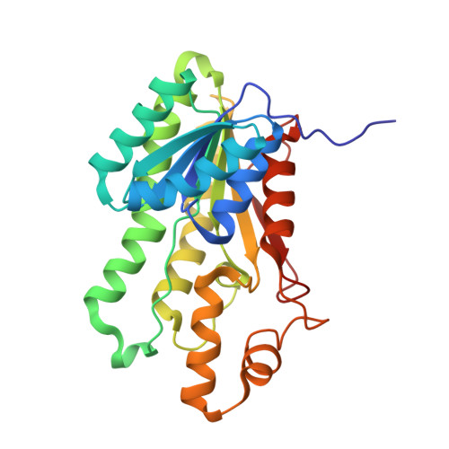
Dissecting the Mechanism of ( R )-3-Hydroxybutyrate Dehydrogenase by Kinetic Isotope Effects, Protein Crystallography, and Computational Chemistry.
Machado, T.F.G., Purg, M., McMahon, S.A., Read, B.J., Oehler, V., Aqvist, J., Gloster, T.M., da Silva, R.G.(2020) ACS Catal 10: 15019-15032
- PubMed: 33391858
- DOI: https://doi.org/10.1021/acscatal.0c04736
- Primary Citation of Related Structures:
6ZZO, 6ZZP, 6ZZQ, 6ZZS - PubMed Abstract:
The enzyme ( R )-3-hydroxybutyrate dehydrogenase (HBDH) catalyzes the enantioselective reduction of 3-oxocarboxylates to ( R )-3-hydroxycarboxylates, the monomeric precursors of biodegradable polyesters. Despite its application in asymmetric reduction, which prompted several engineering attempts of this enzyme, the order of chemical events in the active site, their contributions to limit the reaction rate, and interactions between the enzyme and non-native 3-oxocarboxylates have not been explored. Here, a combination of kinetic isotope effects, protein crystallography, and quantum mechanics/molecular mechanics (QM/MM) calculations were employed to dissect the HBDH mechanism. Initial velocity patterns and primary deuterium kinetic isotope effects establish a steady-state ordered kinetic mechanism for acetoacetate reduction by a psychrophilic and a mesophilic HBDH, where hydride transfer is not rate limiting. Primary deuterium kinetic isotope effects on the reduction of 3-oxovalerate indicate that hydride transfer becomes more rate limiting with this non-native substrate. Solvent and multiple deuterium kinetic isotope effects suggest hydride and proton transfers occur in the same transition state. Crystal structures were solved for both enzymes complexed to NAD + :acetoacetate and NAD + :3-oxovalerate, illustrating the structural basis for the stereochemistry of the 3-hydroxycarboxylate products. QM/MM calculations using the crystal structures as a starting point predicted a higher activation energy for 3-oxovalerate reduction catalyzed by the mesophilic HBDH, in agreement with the higher reaction rate observed experimentally for the psychrophilic orthologue. Both transition states show concerted, albeit not synchronous, proton and hydride transfers to 3-oxovalerate. Setting the MM partial charges to zero results in identical reaction activation energies with both orthologues, suggesting the difference in activation energy between the reactions catalyzed by cold- and warm-adapted HBDHs arises from differential electrostatic stabilization of the transition state. Mutagenesis and phylogenetic analysis reveal the catalytic importance of His150 and Asn145 in the respective orthologues.
- School of Chemistry and School of Biology, Biomedical Sciences Research Complex, University of St Andrews, St Andrews, Fife KY16 9ST, United Kingdom.
Organizational Affiliation:


