6ZZ5

Structure of soluble SmhB of the tripartite alpha-pore forming toxin, Smh, from Serratia marcescens.


Experimental Data Snapshot

  • Method: X-RAY DIFFRACTION
  • Resolution: 1.84 Å
  • R-Value Free: 0.224 
  • R-Value Work: 0.187 

wwPDB Validation   3D Report Full Report


This is version 1.1 of the entry. See complete history


Literature

Characterisation of a tripartite alpha-pore forming toxin from Serratia marcescens

Churchill-Angus, A.M.Schofield, T.H.B.Marlow, T.R.Sedelnikova, S.E.Wilson, J.S.Rafferty, J.B.Baker, P.J.

(2021) Sci Rep 11: 6447

  • DOI: https://doi.org/10.1038/s41598-021-85726-0
  • Primary Citation of Related Structures:  
    6ZZ5, 6ZZH, 7A0G, 7A26, 7A27

  • PubMed Abstract: 

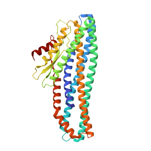
    Tripartite members of the ClyA family of α-PFTs have recently been identified in a number of pathogenic Gram-negative bacteria, including the human pathogen Serratia marcescens. Structures of a Gram-negative A component and a tripartite α-PFT complete pore are unknown and a mechanism for pore formation is still uncertain. Here we characterise the tripartite SmhABC toxin from S. marcescens and propose a mechanism of pore assembly. We present the structure of soluble SmhA, as well as the soluble and pore forms of SmhB. We show that the β-tongue soluble structure is well conserved in the family and propose two conserved latches between the head and tail domains that are broken on the soluble to pore conformational change. Using the structures of individual components, sequence analysis and docking predictions we illustrate how the A, B and C protomers would assemble on the membrane to produce a complete tripartite α-PFT pore.


  • Organizational Affiliation

    Department of Molecular Biology and Biotechnology, University of Sheffield, Firth Court, Western Bank, Sheffield, S10 2TN, South Yorkshire, UK.


Macromolecules
Find similar proteins by:  (by identity cutoff)  |  3D Structure
Entity ID: 1
MoleculeChains Sequence LengthOrganismDetailsImage
SmhBA [auth AAA],
B [auth BBB]
366Serratia marcescensMutation(s): 0 
Gene Names: BHU62_20105
Membrane Entity: Yes 
UniProt
Find proteins for A0A1Q4NVM7 (Serratia marcescens)
Explore A0A1Q4NVM7 
Go to UniProtKB:  A0A1Q4NVM7
Entity Groups  
Sequence Clusters30% Identity50% Identity70% Identity90% Identity95% Identity100% Identity
UniProt GroupA0A1Q4NVM7
Sequence Annotations
Expand
  • Reference Sequence
Small Molecules
Ligands 2 Unique
IDChains Name / Formula / InChI Key2D Diagram3D Interactions
SO4
Query on SO4

Download Ideal Coordinates CCD File 
C [auth AAA]
D [auth AAA]
E [auth AAA]
F [auth AAA]
G [auth AAA]
C [auth AAA],
D [auth AAA],
E [auth AAA],
F [auth AAA],
G [auth AAA],
L [auth BBB],
M [auth BBB],
N [auth BBB]
SULFATE ION
O4 S
QAOWNCQODCNURD-UHFFFAOYSA-L
EDO
Query on EDO

Download Ideal Coordinates CCD File 
H [auth AAA]
I [auth AAA]
J [auth AAA]
K [auth AAA]
O [auth BBB]
H [auth AAA],
I [auth AAA],
J [auth AAA],
K [auth AAA],
O [auth BBB],
P [auth BBB],
Q [auth BBB]
1,2-ETHANEDIOL
C2 H6 O2
LYCAIKOWRPUZTN-UHFFFAOYSA-N
Experimental Data & Validation

Experimental Data

  • Method: X-RAY DIFFRACTION
  • Resolution: 1.84 Å
  • R-Value Free: 0.224 
  • R-Value Work: 0.187 
  • Space Group: P 21 21 21
Unit Cell:
Length ( Å )Angle ( ˚ )
a = 50.94α = 90
b = 113.95β = 90
c = 130.37γ = 90
Software Package:
Software NamePurpose
REFMACrefinement
xia2data reduction
xia2data scaling
PHASERphasing

Structure Validation

View Full Validation Report


Entry History 

Deposition Data

Revision History  (Full details and data files)

  • Version 1.0: 2021-03-31
    Type: Initial release
  • Version 1.1: 2024-01-31
    Changes: Data collection, Database references, Derived calculations, Refinement description