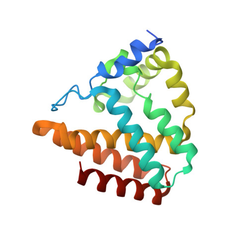
How cyanophage S-2L rejects adenine and incorporates 2-aminoadenine to saturate hydrogen bonding in its DNA.
Czernecki, D., Legrand, P., Tekpinar, M., Rosario, S., Kaminski, P.A., Delarue, M.(2021) Nat Commun 12: 2420-2420
- PubMed: 33893297
- DOI: https://doi.org/10.1038/s41467-021-22626-x
- Primary Citation of Related Structures:
6ZP9, 6ZPA, 6ZPB, 6ZPC - PubMed Abstract:
Bacteriophages have long been known to use modified bases in their DNA to prevent cleavage by the host's restriction endonucleases. Among them, cyanophage S-2L is unique because its genome has all its adenines (A) systematically replaced by 2-aminoadenines (Z). Here, we identify a member of the PrimPol family as the sole possible polymerase of S-2L and we find it can incorporate both A and Z in front of a T. Its crystal structure at 1.5 Å resolution confirms that there is no structural element in the active site that could lead to the rejection of A in front of T. To resolve this contradiction, we show that a nearby gene is a triphosphohydolase specific of dATP (DatZ), that leaves intact all other dNTPs, including dZTP. This explains the absence of A in S-2L genome. Crystal structures of DatZ with various ligands, including one at sub-angstrom resolution, allow to describe its mechanism as a typical two-metal-ion mechanism and to set the stage for its engineering.
- Unit of Structural Dynamics of Biological Macromolecules, CNRS UMR 3528, 25-28 rue du Docteur Roux, Institut Pasteur, Paris, France.
Organizational Affiliation:


