6ZMS | pdb_00006zms

Coxsackievirus B4 strain E2


Experimental Data Snapshot

  • Method: ELECTRON MICROSCOPY
  • Resolution: 3.40 Å
  • Aggregation State: PARTICLE 
  • Reconstruction Method: SINGLE PARTICLE 

wwPDB Validation 3D Report Full Report

Validation slider image for 6ZMS

This is version 2.0 of the entry. See complete history

Literature

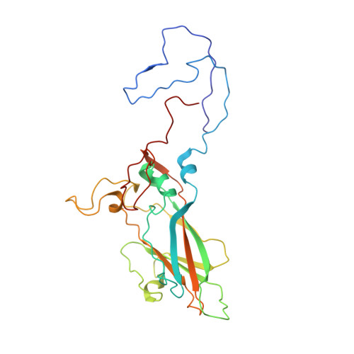
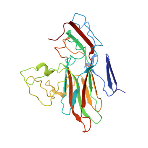
Identification of a conserved virion-stabilizing network inside the interprotomer pocket of enteroviruses.

Flatt, J.W.Domanska, A.Seppala, A.L.Butcher, S.J.

(2021) Commun Biol 4: 250-250

  • DOI: https://doi.org/10.1038/s42003-021-01779-x
  • Primary Citation Related Structures: 
    6ZCK, 6ZCL, 6ZMS

  • PubMed Abstract: 

    Enteroviruses pose a persistent and widespread threat to human physical health, with no specific treatments available. Small molecule capsid binders have the potential to be developed as antivirals that prevent virus attachment and entry into host cells. To aid with broad-range drug development, we report here structures of coxsackieviruses B3 and B4 bound to different interprotomer-targeting capsid binders using single-particle cryo-EM. The EM density maps are beyond 3 Å resolution, providing detailed information about interactions in the ligand-binding pocket. Comparative analysis revealed the residues that form a conserved virion-stabilizing network at the interprotomer site, and showed the small molecule properties that allow anchoring in the pocket to inhibit virus disassembly.


  • Organizational Affiliation
    • Faculty of Biological and Environmental Sciences, Molecular and Integrative Bioscience Research Programme, University of Helsinki, Helsinki, Finland.

Macromolecule Content 

  • Total Structure Weight: 92.34 kDa 
  • Atom Count: 6,396 
  • Modeled Residue Count: 818 
  • Deposited Residue Count: 830 
  • Unique protein chains: 4

Macromolecules

Find similar proteins by:|  3D Structure
Entity ID: 1
MoleculeChains  Sequence LengthOrganismDetailsImage
Capsid protein VP1271Coxsackievirus B4 (strain E2)Mutation(s): 5 
UniProt
Find proteins for Q86887 (Coxsackievirus B4 (strain E2))
Explore Q86887 
Go to UniProtKB:  Q86887
Entity Groups
Sequence Clusters30% Identity50% Identity70% Identity90% Identity95% Identity100% Identity
UniProt GroupQ86887
Sequence Annotations
Expand
Reference Sequence
Find similar proteins by:|  3D Structure
Entity ID: 2
MoleculeChains  Sequence LengthOrganismDetailsImage
Capsid protein VP2252Coxsackievirus B4 (strain E2)Mutation(s): 1 
UniProt
Find proteins for Q86887 (Coxsackievirus B4 (strain E2))
Explore Q86887 
Go to UniProtKB:  Q86887
Entity Groups
Sequence Clusters30% Identity50% Identity70% Identity90% Identity95% Identity100% Identity
UniProt GroupQ86887
Sequence Annotations
Expand
Reference Sequence
Find similar proteins by:|  3D Structure
Entity ID: 3
MoleculeChains  Sequence LengthOrganismDetailsImage
Capsid protein VP3238Coxsackievirus B4 (strain E2)Mutation(s): 0 
UniProt
Find proteins for Q86887 (Coxsackievirus B4 (strain E2))
Explore Q86887 
Go to UniProtKB:  Q86887
Entity Groups
Sequence Clusters30% Identity50% Identity70% Identity90% Identity95% Identity100% Identity
UniProt GroupQ86887
Sequence Annotations
Expand
Reference Sequence
Find similar proteins by:|  3D Structure
Entity ID: 4
MoleculeChains  Sequence LengthOrganismDetailsImage
Capsid protein VP469Coxsackievirus B4 (strain E2)Mutation(s): 1 
UniProt
Find proteins for Q86887 (Coxsackievirus B4 (strain E2))
Explore Q86887 
Go to UniProtKB:  Q86887
Entity Groups
Sequence Clusters30% Identity50% Identity70% Identity90% Identity95% Identity100% Identity
UniProt GroupQ86887
Sequence Annotations
Expand
Reference Sequence

Experimental Data & Validation

Experimental Data

  • Method: ELECTRON MICROSCOPY
  • Resolution: 3.40 Å
  • Aggregation State: PARTICLE 
  • Reconstruction Method: SINGLE PARTICLE 

Structure Validation

View Full Validation Report


Entry History 

& Funding Information

Deposition Data


Funding OrganizationLocationGrant Number
Academy of FinlandFinland315950
Sigrid Juselius FoundationFinland--

Revision History  (Full details and data files)

  • Version 1.0: 2021-03-17
    Type: Initial release
  • Version 2.0: 2024-05-01
    Changes: Atomic model, Data collection, Database references, Derived calculations, Source and taxonomy, Structure summary