Structure and mechanism of a phage-encoded SAM lyase revises catalytic function of enzyme family.
Guo, X., Soderholm, A., Kanchugal P, S., Isaksen, G.V., Warsi, O., Eckhard, U., Triguis, S., Gogoll, A., Jerlstrom-Hultqvist, J., Aqvist, J., Andersson, D.I., Selmer, M.(2021) Elife 10
- PubMed: 33567250 Search on PubMedSearch on PubMed Central
- DOI: https://doi.org/10.7554/eLife.61818
- Primary Citation Related Structures:
6ZM9, 6ZMG, 6ZNB - PubMed Abstract:
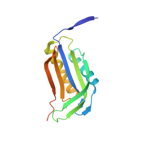
The first S-adenosyl methionine (SAM) degrading enzyme (SAMase) was discovered in bacteriophage T3, as a counter-defense against the bacterial restriction-modification system, and annotated as a SAM hydrolase forming 5'-methyl-thioadenosine (MTA) and L-homoserine. From environmental phages, we recently discovered three SAMases with barely detectable sequence similarity to T3 SAMase and without homology to proteins of known structure. Here, we present the very first phage SAMase structures, in complex with a substrate analogue and the product MTA. The structure shows a trimer of alpha-beta sandwiches similar to the GlnB-like superfamily, with active sites formed at the trimer interfaces. Quantum-mechanical calculations, thin-layer chromatography, and nuclear magnetic resonance spectroscopy demonstrate that this family of enzymes are not hydrolases but lyases forming MTA and L-homoserine lactone in a unimolecular reaction mechanism. Sequence analysis and in vitro and in vivo mutagenesis support that T3 SAMase belongs to the same structural family and utilizes the same reaction mechanism.
- Department of Cell and Molecular Biology, Uppsala University, Uppsala, Sweden.
Organizational Affiliation: