6ZMA | pdb_00006zma

Structure of the tRNA-Monooxygenase enzyme MiaE frozen under 140 bar of krypton using the soak and freeze methodology


Experimental Data Snapshot

  • Method: X-RAY DIFFRACTION
  • Resolution: 2.15 Å
  • R-Value Free: 
    0.244 (Depositor), 0.246 (DCC) 
  • R-Value Work: 
    0.180 (Depositor), 0.189 (DCC) 
  • R-Value Observed: 
    0.183 (Depositor) 

Starting Model: experimental
View more details

wwPDB Validation 3D Report Full Report

Validation slider image for 6ZMA

This is version 1.2 of the entry. See complete history

Literature

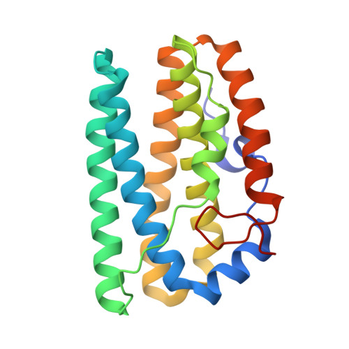
Structural, biochemical and functional analyses of tRNA-monooxygenase enzyme MiaE from Pseudomonas putida provide insights into tRNA/MiaE interaction.

Carpentier, P.Lepretre, C.Basset, C.Douki, T.Torelli, S.Duarte, V.Hamdane, D.Fontecave, M.Atta, M.

(2020) Nucleic Acids Res 48: 9918-9930

  • DOI: https://doi.org/10.1093/nar/gkaa667
  • Primary Citation Related Structures: 
    6ZMA, 6ZMB, 6ZMC

  • PubMed Abstract: 

    MiaE (2-methylthio-N6-isopentenyl-adenosine37-tRNA monooxygenase) is a unique non-heme diiron enzyme that catalyzes the O2-dependent post-transcriptional allylic hydroxylation of a hypermodified nucleotide 2-methylthio-N6-isopentenyl-adenosine (ms2i6A37) at position 37 of selected tRNA molecules to produce 2-methylthio-N6-4-hydroxyisopentenyl-adenosine (ms2io6A37). Here, we report the in vivo activity, biochemical, spectroscopic characterization and X-ray crystal structure of MiaE from Pseudomonas putida. The investigation demonstrates that the putative pp-2188 gene encodes a MiaE enzyme. The structure shows that Pp-MiaE consists of a catalytic diiron(III) domain with a four alpha-helix bundle fold. A docking model of Pp-MiaE in complex with tRNA, combined with site directed mutagenesis and in vivo activity shed light on the importance of an additional linker region for substrate tRNA recognition. Finally, krypton-pressurized Pp-MiaE experiments, revealed the presence of defined O2 site along a conserved hydrophobic tunnel leading to the diiron active center.


  • Organizational Affiliation
    • Univ. Grenoble Alpes, CEA, CNRS, CBM-UMR 5249, 17 avenue des martyrs, Grenoble, France.

Macromolecule Content 

  • Total Structure Weight: 48.45 kDa 
  • Atom Count: 3,483 
  • Modeled Residue Count: 398 
  • Deposited Residue Count: 410 
  • Unique protein chains: 1

Macromolecules

Find similar proteins by:|  3D Structure
Entity ID: 1
MoleculeChains  Sequence LengthOrganismDetailsImage
tRNA hydroxylaseA [auth B],
B [auth C]
205Pseudomonas putidaMutation(s): 0 
Gene Names: AYO08_21560CBL13_00280CBP06_18695
UniProt
Find proteins for A0A179QS89 (Pseudomonas putida)
Explore A0A179QS89 
Go to UniProtKB:  A0A179QS89
Entity Groups
Sequence Clusters30% Identity50% Identity70% Identity90% Identity95% Identity100% Identity
UniProt GroupA0A179QS89
Sequence Annotations
Expand
Reference Sequence

Small Molecules

Ligands 8 Unique
IDChains Name / Formula / InChI Key2D Diagram3D Interactions
PG4

Query on PG4


Download:Ideal Coordinates CCD File
L [auth B]TETRAETHYLENE GLYCOL
C8 H18 O5
UWHCKJMYHZGTIT-UHFFFAOYSA-N
TRS

Query on TRS


Download:Ideal Coordinates CCD File
H [auth B],
R [auth C]
2-AMINO-2-HYDROXYMETHYL-PROPANE-1,3-DIOL
C4 H12 N O3
LENZDBCJOHFCAS-UHFFFAOYSA-O
PEG

Query on PEG


Download:Ideal Coordinates CCD File
I [auth B]
J [auth B]
K [auth B]
S [auth C]
T [auth C]
I [auth B],
J [auth B],
K [auth B],
S [auth C],
T [auth C],
U [auth C],
V [auth C]
DI(HYDROXYETHYL)ETHER
C4 H10 O3
MTHSVFCYNBDYFN-UHFFFAOYSA-N
KR
(Subject of Investigation/LOI)

Query on KR


Download:Ideal Coordinates CCD File
M [auth B]
N [auth B]
W [auth C]
X [auth C]
Y [auth C]
M [auth B],
N [auth B],
W [auth C],
X [auth C],
Y [auth C],
Z [auth C]
KRYPTON
Kr
DNNSSWSSYDEUBZ-UHFFFAOYSA-N
EDO

Query on EDO


Download:Ideal Coordinates CCD File
AA [auth C],
BA [auth C],
O [auth B]
1,2-ETHANEDIOL
C2 H6 O2
LYCAIKOWRPUZTN-UHFFFAOYSA-N
FE
(Subject of Investigation/LOI)

Query on FE


Download:Ideal Coordinates CCD File
C [auth B],
D [auth B],
P [auth C],
Q [auth C]
FE (III) ION
Fe
VTLYFUHAOXGGBS-UHFFFAOYSA-N
CA

Query on CA


Download:Ideal Coordinates CCD File
F [auth B],
G [auth B]
CALCIUM ION
Ca
BHPQYMZQTOCNFJ-UHFFFAOYSA-N
CL

Query on CL


Download:Ideal Coordinates CCD File
E [auth B]CHLORIDE ION
Cl
VEXZGXHMUGYJMC-UHFFFAOYSA-M

Experimental Data & Validation

Experimental Data

  • Method: X-RAY DIFFRACTION
  • Resolution: 2.15 Å
  • R-Value Free:  0.244 (Depositor), 0.246 (DCC) 
  • R-Value Work:  0.180 (Depositor), 0.189 (DCC) 
  • R-Value Observed: 0.183 (Depositor) 
Space Group: C 1 2 1
Unit Cell:
Length ( Å )Angle ( ˚ )
a = 117.622α = 90
b = 52.3β = 90.74
c = 79.407γ = 90
Software Package:
Software NamePurpose
REFMACrefinement
PDB_EXTRACTdata extraction
XDSdata reduction
XSCALEdata scaling
PHASERphasing

Structure Validation

View Full Validation Report


Entry History 

& Funding Information

Deposition Data


Funding OrganizationLocationGrant Number
French National Research AgencyFranceANR-15-CE21-0020

Revision History  (Full details and data files)

  • Version 1.0: 2020-09-02
    Type: Initial release
  • Version 1.1: 2020-10-07
    Changes: Database references
  • Version 1.2: 2024-01-31
    Changes: Advisory, Data collection, Database references, Refinement description