6ZL4 | pdb_00006zl4

the structure of glutamate transporter homologue GltTk in complex with the photo switchable compound (cis)


Experimental Data Snapshot

  • Method: X-RAY DIFFRACTION
  • Resolution: 3.00 Å
  • R-Value Free: 
    0.288 (Depositor), 0.291 (DCC) 
  • R-Value Work: 
    0.255 (Depositor), 0.261 (DCC) 
  • R-Value Observed: 
    0.256 (Depositor) 

Starting Model: experimental
View more details

wwPDB Validation 3D Report Full Report

Validation slider image for 6ZL4

Ligand Structure Quality Assessment 


This is version 1.2 of the entry. See complete history

Literature

Structural Aspects of Photopharmacology: Insight into the Binding of Photoswitchable and Photocaged Inhibitors to the Glutamate Transporter Homologue.

Arkhipova, V.Fu, H.Hoorens, M.W.H.Trinco, G.Lameijer, L.N.Marin, E.Feringa, B.L.Poelarends, G.J.Szymanski, W.Slotboom, D.J.Guskov, A.

(2021) J Am Chem Soc 143: 1513-1520

  • DOI: https://doi.org/10.1021/jacs.0c11336
  • Primary Citation Related Structures: 
    6ZGB, 6ZL4, 6ZLH

  • PubMed Abstract: 

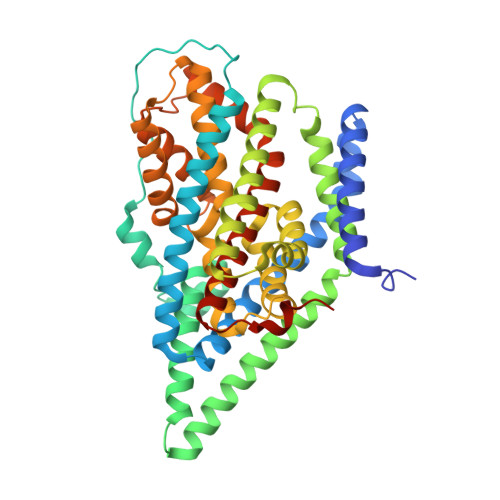
    Photopharmacology addresses the challenge of drug selectivity and side effects through creation of photoresponsive molecules activated with light with high spatiotemporal precision. This is achieved through incorporation of molecular photoswitches and photocages into the pharmacophore. However, the structural basis for the light-induced modulation of inhibitory potency in general is still missing, which poses a major design challenge for this emerging field of research. Here we solved crystal structures of the glutamate transporter homologue Glt Tk in complex with photoresponsive transport inhibitors-azobenzene derivative of TBOA (both in trans and cis configuration) and with the photocaged compound ONB-hydroxyaspartate. The essential role of glutamate transporters in the functioning of the central nervous system renders them potential therapeutic targets in the treatment of neurodegenerative diseases. The obtained structures provide a clear structural insight into the origins of photocontrol in photopharmacology and lay the foundation for application of photocontrolled ligands to study the transporter dynamics by using time-resolved X-ray crystallography.


  • Organizational Affiliation
    • University Medical Center Groningen, Department of Radiology, Medical Imaging Center, University of Groningen, Hanzeplein 1, 9713 GZ Groningen, The Netherlands.

Macromolecule Content 

  • Total Structure Weight: 146.04 kDa 
  • Atom Count: 9,881 
  • Modeled Residue Count: 1,270 
  • Deposited Residue Count: 1,314 
  • Unique protein chains: 1

Macromolecules

Find similar proteins by:|  3D Structure
Entity ID: 1
MoleculeChains  Sequence LengthOrganismDetailsImage
Proton/glutamate symporter, SDF family
A, B, C
438Thermococcus kodakarensisMutation(s): 0 
Gene Names: TK0986
Membrane Entity: Yes 
UniProt
Find proteins for Q5JID0 (Thermococcus kodakarensis (strain ATCC BAA-918 / JCM 12380 / KOD1))
Explore Q5JID0 
Go to UniProtKB:  Q5JID0
Entity Groups
Sequence Clusters30% Identity50% Identity70% Identity90% Identity95% Identity100% Identity
UniProt GroupQ5JID0
Sequence Annotations
Expand
Reference Sequence

Small Molecules

Ligands 7 Unique
IDChains Name / Formula / InChI Key2D Diagram3D Interactions
DMU

Query on DMU


Download:Ideal Coordinates CCD File
DA [auth C],
E [auth A],
T [auth B]
DECYL-BETA-D-MALTOPYRANOSIDE
C22 H42 O11
WOQQAWHSKSSAGF-WXFJLFHKSA-N
QM5

Query on QM5


Download:Ideal Coordinates CCD File
CA [auth C],
D [auth A],
P [auth B]
(2~{S},3~{S})-2-azanyl-3-[[4-[2-(4-methoxyphenyl)hydrazinyl]phenyl]methoxy]butanedioic acid
C18 H21 N3 O6
FVCUHYYDCQQINH-HOTGVXAUSA-N
1PE

Query on 1PE


Download:Ideal Coordinates CCD File
FA [auth C]PENTAETHYLENE GLYCOL
C10 H22 O6
JLFNLZLINWHATN-UHFFFAOYSA-N
PG4

Query on PG4


Download:Ideal Coordinates CCD File
F [auth A],
G [auth A],
I [auth A]
TETRAETHYLENE GLYCOL
C8 H18 O5
UWHCKJMYHZGTIT-UHFFFAOYSA-N
PGE

Query on PGE


Download:Ideal Coordinates CCD File
EA [auth C]
H [auth A]
J [auth A]
Q [auth B]
U [auth B]
EA [auth C],
H [auth A],
J [auth A],
Q [auth B],
U [auth B],
V [auth B]
TRIETHYLENE GLYCOL
C6 H14 O4
ZIBGPFATKBEMQZ-UHFFFAOYSA-N
PEG

Query on PEG


Download:Ideal Coordinates CCD File
AA [auth B]
BA [auth B]
JA [auth C]
KA [auth C]
LA [auth C]
AA [auth B],
BA [auth B],
JA [auth C],
KA [auth C],
LA [auth C],
MA [auth C],
N [auth A],
NA [auth C],
O [auth A],
OA [auth C],
PA [auth C],
R [auth B],
S [auth B],
Z [auth B]
DI(HYDROXYETHYL)ETHER
C4 H10 O3
MTHSVFCYNBDYFN-UHFFFAOYSA-N
NA

Query on NA


Download:Ideal Coordinates CCD File
GA [auth C]
HA [auth C]
IA [auth C]
K [auth A]
L [auth A]
GA [auth C],
HA [auth C],
IA [auth C],
K [auth A],
L [auth A],
M [auth A],
W [auth B],
X [auth B],
Y [auth B]
SODIUM ION
Na
FKNQFGJONOIPTF-UHFFFAOYSA-N

Experimental Data & Validation

Experimental Data

  • Method: X-RAY DIFFRACTION
  • Resolution: 3.00 Å
  • R-Value Free:  0.288 (Depositor), 0.291 (DCC) 
  • R-Value Work:  0.255 (Depositor), 0.261 (DCC) 
  • R-Value Observed: 0.256 (Depositor) 
Space Group: P 32 2 1
Unit Cell:
Length ( Å )Angle ( ˚ )
a = 117.21α = 90
b = 117.21β = 90
c = 309.41γ = 120
Software Package:
Software NamePurpose
XSCALEdata scaling
PHENIXrefinement
PDB_EXTRACTdata extraction
XSCALEdata reduction
PHASERphasing

Structure Validation

View Full Validation Report


Ligand Structure Quality Assessment 


Entry History 

& Funding Information

Deposition Data


Funding OrganizationLocationGrant Number
Netherlands Organisation for Scientific Research (NWO)Netherlands--

Revision History  (Full details and data files)

  • Version 1.0: 2021-01-27
    Type: Initial release
  • Version 1.1: 2021-02-03
    Changes: Database references
  • Version 1.2: 2024-01-31
    Changes: Data collection, Database references, Refinement description