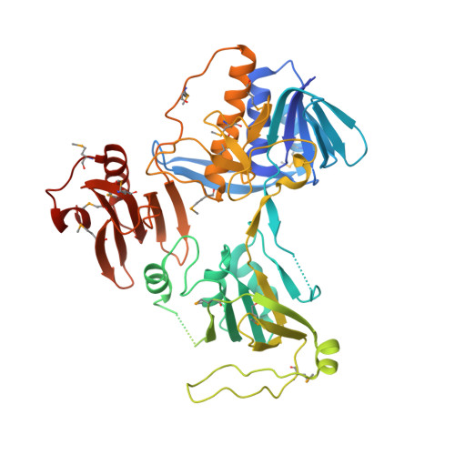
The tRNA ligase complex (tRNA-LC) splices precursor tRNAs (pre-tRNA), and Xbp1-mRNA during the unfolded protein response (UPR). In aerobic conditions, a cysteine residue bound to two metal ions in its ancient, catalytic subunit RTCB could make the tRNA-LC susceptible to oxidative inactivation. Here, we confirm this hypothesis and reveal a co-evolutionary association between the tRNA-LC and PYROXD1, a conserved and essential oxidoreductase. We reveal that PYROXD1 preserves the activity of the mammalian tRNA-LC in pre-tRNA splicing and UPR. PYROXD1 binds the tRNA-LC in the presence of NAD(P)H and converts RTCB-bound NAD(P)H into NAD(P) + , a typical oxidative co-enzyme. However, NAD(P) + here acts as an antioxidant and protects the tRNA-LC from oxidative inactivation, which is dependent on copper ions. Genetic variants of PYROXD1 that cause human myopathies only partially support tRNA-LC activity. Thus, we establish the tRNA-LC as an oxidation-sensitive metalloenzyme, safeguarded by the flavoprotein PYROXD1 through an unexpected redox mechanism.
Organizational Affiliation:
Max Perutz Labs, Medical University of Vienna, Vienna BioCenter (VBC), Dr. Bohr-Gasse 9/2, 1030 Vienna, Austria.
Institute of Biochemistry, Graz University of Technology, Petersgasse 12/2, 8010 Graz, Austria; Department of Medical Biochemistry and Biophysics, Karolinska Institutet, 171 77 Stockholm, Sweden.
Department of Biochemistry, University of Zurich, Winterthurerstrasse 190, 8057 Zurich, Switzerland.
Research Institute of Molecular Pathology (IMP), Vienna BioCenter (VBC), Campus-Vienna-BioCenter 1, 1030 Vienna, Austria.
Institute of Molecular Biotechnology of the Austrian Academy of Sciences (IMBA), Vienna BioCenter (VBC), Dr. Bohr-Gasse 3, 1030 Vienna, Austria; AnnJi Pharmaceutical, Taipei, Taiwan.
Department of Pharmaceutical Chemistry, Faculty of Pharmacy, University of Belgrade, Vojvode Stepe 450, 11221 Belgrade, Serbia.
Max Perutz Labs, University of Vienna, Vienna BioCenter (VBC), Dr. Bohr-Gasse 9, 1030 Vienna, Austria.
Institute of Molecular Biotechnology of the Austrian Academy of Sciences (IMBA), Vienna BioCenter (VBC), Dr. Bohr-Gasse 3, 1030 Vienna, Austria; Department of Internal Medicine III (Cardiology and Angiology), Medical University of Innsbruck, Anichstraße 35, 6020 Innsbruck, Austria.
Research Institute of Molecular Pathology (IMP), Vienna BioCenter (VBC), Campus-Vienna-BioCenter 1, 1030 Vienna, Austria; Sir William Dunn School of Pathology, University of Oxford, South Parks Road, OX1 3RE Oxford, UK.
Institute of Molecular and Cell Biology, University of Tartu, Riia 23, 51010 Tartu, Estonia.
Research Institute of Molecular Pathology (IMP), Vienna BioCenter (VBC), Campus-Vienna-BioCenter 1, 1030 Vienna, Austria; Department of Pure and Applied Chemistry, University of Strathclyde, 295 Cathedral Street, G1 1XL Glasgow, UK.
Research Institute of Molecular Pathology (IMP), Vienna BioCenter (VBC), Campus-Vienna-BioCenter 1, 1030 Vienna, Austria; Institute of Molecular Biotechnology of the Austrian Academy of Sciences (IMBA), Vienna BioCenter (VBC), Dr. Bohr-Gasse 3, 1030 Vienna, Austria.
Institute of Molecular Biotechnology of the Austrian Academy of Sciences (IMBA), Vienna BioCenter (VBC), Dr. Bohr-Gasse 3, 1030 Vienna, Austria; Department of Medical Genetics, Life Science Institute, University of British Columbia, C201 - 4500 Oak Street, V6H 3N1 Vancouver, BC, Canada.
Institute of Biochemistry, Graz University of Technology, Petersgasse 12/2, 8010 Graz, Austria.
Max Perutz Labs, Medical University of Vienna, Vienna BioCenter (VBC), Dr. Bohr-Gasse 9/2, 1030 Vienna, Austria. Electronic address: stefan.weitzer@meduniwien.ac.at.
Max Perutz Labs, Medical University of Vienna, Vienna BioCenter (VBC), Dr. Bohr-Gasse 9/2, 1030 Vienna, Austria. Electronic address: javier.martinez@meduniwien.ac.at.