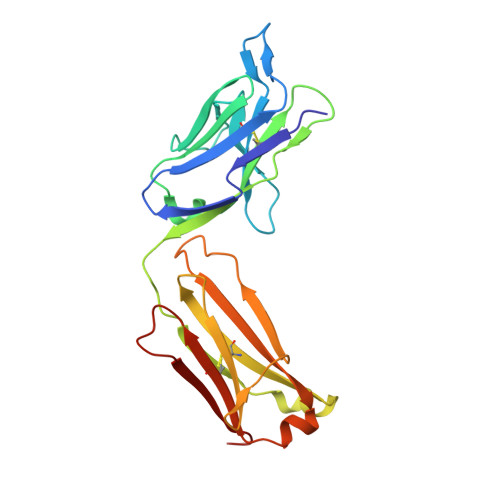
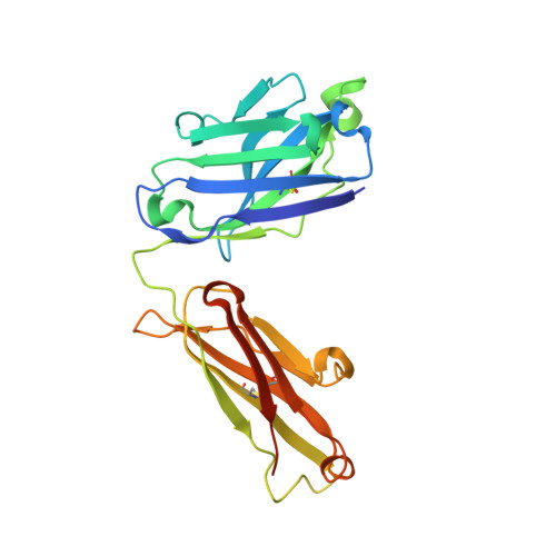
Insertion of atypical glycans into the tumor antigen-binding site identifies DLBCLs with distinct origin and behavior.
Chiodin, G., Allen, J.D., Bryant, D.J., Rock, P., Martino, E.A., Valle-Argos, B., Duriez, P.J., Watanabe, Y., Henderson, I., Blachly, J.S., McCann, K.J., Strefford, J.C., Packham, G., Geijtenbeek, T.B.H., Figdor, C.G., Wright, G.W., Staudt, L.M., Burack, R., Bowden, T.A., Crispin, M., Stevenson, F.K., Forconi, F.(2021) Blood 138: 1570-1582
- PubMed: 34424958 Search on PubMedSearch on PubMed Central
- DOI: https://doi.org/10.1182/blood.2021012052
- Primary Citation Related Structures:
6ZEC - PubMed Abstract:
Glycosylation of the surface immunoglobulin (Ig) variable region is a remarkable follicular lymphoma-associated feature rarely seen in normal B cells. Here, we define a subset of diffuse large B-cell lymphomas (DLBCLs) that acquire N-glycosylation sites selectively in the Ig complementarity-determining regions (CDRs) of the antigen-binding sites. Mass spectrometry and X-ray crystallography demonstrate how the inserted glycans are stalled at oligomannose-type structures because they are buried in the CDR loops. Acquisition of sites occurs in ∼50% of germinal-center B-cell-like DLBCL (GCB-DLBCL), mainly of the genetic EZB subtype, irrespective of IGHV-D-J use. This markedly contrasts with the activated B-cell-like DLBCL Ig, which rarely has sites in the CDR and does not seem to acquire oligomannose-type structures. Acquisition of CDR-located acceptor sites associates with mutations of epigenetic regulators and BCL2 translocations, indicating an origin shared with follicular lymphoma. Within the EZB subtype, these sites are associated with more rapid disease progression and with significant gene set enrichment of the B-cell receptor, PI3K/AKT/MTORC1 pathway, glucose metabolism, and MYC signaling pathways, particularly in the fraction devoid of MYC translocations. The oligomannose-type glycans on the lymphoma cells interact with the candidate lectin dendritic cell-specific intercellular adhesion molecule 3 grabbing non-integrin (DC-SIGN), mediating low-level signals, and lectin-expressing cells form clusters with lymphoma cells. Both clustering and signaling are inhibited by antibodies specifically targeting the DC-SIGN carbohydrate recognition domain. Oligomannosylation of the tumor Ig is a posttranslational modification that readily identifies a distinct GCB-DLBCL category with more aggressive clinical behavior, and it could be a potential precise therapeutic target via antibody-mediated inhibition of the tumor Ig interaction with DC-SIGN-expressing M2-polarized macrophages.
- School of Cancer Sciences, Cancer Research United Kingdom Southampton Centre, Faculty of Medicine.
Organizational Affiliation: