6ZCJ

14-3-3sigma in complex with SLP76pS376 phosphopeptide crystal structure


Experimental Data Snapshot

  • Method: X-RAY DIFFRACTION
  • Resolution: 1.53 Å
  • R-Value Free: 0.190 
  • R-Value Work: 0.168 
  • R-Value Observed: 0.169 

wwPDB Validation   3D Report Full Report


This is version 1.2 of the entry. See complete history


Literature

The 14-3-3/SLP76 protein-protein interaction in T-cell receptor signalling: a structural and biophysical characterization.

Soini, L.Leysen, S.Davis, J.Westwood, M.Ottmann, C.

(2021) FEBS Lett 595: 404-414

  • DOI: https://doi.org/10.1002/1873-3468.13993
  • Primary Citation of Related Structures:  
    6ZCJ

  • PubMed Abstract: 

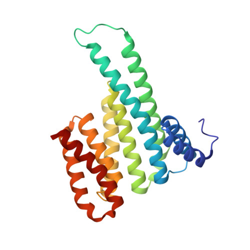
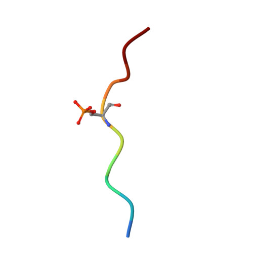
    The SH2 domain-containing protein of 76 kDa, SLP76, is an important adaptor protein that coordinates a complex protein network downstream of T-cell receptors (TCR), ultimately regulating the immune response. Upon phosphorylation on Ser376, SLP76 interacts with 14-3-3 adaptor proteins, which leads to its proteolytic degradation. This provides a negative feedback mechanism by which TCR signalling can be controlled. To gain insight into the 14-3-3/SLP76 protein-protein interaction (PPI), we have determined a high-resolution crystal structure of a SLP76 synthetic peptide containing Ser376 with 14-3-3σ. We then characterized its binding to 14-3-3 proteins biophysically by means of fluorescence polarization and isothermal titration calorimetry. Furthermore, we generated two recombinant SLP76 protein constructs and characterized their binding to 14-3-3. Our work lays the foundation for drug design efforts aimed at targeting the 14-3-3/SLP76 interaction and, thereby, TCR signalling.


  • Organizational Affiliation

    Laboratory of Chemical Biology, Department of Biomedical Engineering and Institute for Complex Molecular Systems, Eindhoven University of Technology, Eindhoven, the Netherlands.


Macromolecules
Find similar proteins by:  (by identity cutoff)  |  3D Structure
Entity ID: 1
MoleculeChains Sequence LengthOrganismDetailsImage
14-3-3 protein sigma236Homo sapiensMutation(s): 0 
Gene Names: SFNHME1
UniProt & NIH Common Fund Data Resources
Find proteins for P31947 (Homo sapiens)
Explore P31947 
Go to UniProtKB:  P31947
PHAROS:  P31947
GTEx:  ENSG00000175793 
Entity Groups  
Sequence Clusters30% Identity50% Identity70% Identity90% Identity95% Identity100% Identity
UniProt GroupP31947
Sequence Annotations
Expand
  • Reference Sequence

Find similar proteins by:  Sequence   |   3D Structure  

Entity ID: 2
MoleculeChains Sequence LengthOrganismDetailsImage
SLP76pS376B [auth P]10synthetic constructMutation(s): 0 
UniProt & NIH Common Fund Data Resources
Find proteins for Q13094 (Homo sapiens)
Explore Q13094 
Go to UniProtKB:  Q13094
PHAROS:  Q13094
GTEx:  ENSG00000043462 
Entity Groups  
Sequence Clusters30% Identity50% Identity70% Identity90% Identity95% Identity100% Identity
UniProt GroupQ13094
Sequence Annotations
Expand
  • Reference Sequence
Experimental Data & Validation

Experimental Data

  • Method: X-RAY DIFFRACTION
  • Resolution: 1.53 Å
  • R-Value Free: 0.190 
  • R-Value Work: 0.168 
  • R-Value Observed: 0.169 
  • Space Group: C 2 2 21
Unit Cell:
Length ( Å )Angle ( ˚ )
a = 82.098α = 90
b = 111.876β = 90
c = 62.4γ = 90
Software Package:
Software NamePurpose
XDSdata scaling
XDSdata reduction
PHASERphasing
PHENIXrefinement
Cootmodel building

Structure Validation

View Full Validation Report


Entry History & Funding Information

Deposition Data


Funding OrganizationLocationGrant Number
European CommissionNetherlands675179

Revision History  (Full details and data files)

  • Version 1.0: 2020-12-02
    Type: Initial release
  • Version 1.1: 2021-06-16
    Changes: Database references
  • Version 1.2: 2024-01-24
    Changes: Data collection, Database references, Refinement description