6Z6I | pdb_00006z6i

SARS-CoV-2 Macrodomain in complex with ADP-HPD


Experimental Data Snapshot

  • Method: X-RAY DIFFRACTION
  • Resolution: 2.00 Å
  • R-Value Free: 
    0.279 (Depositor), 0.283 (DCC) 
  • R-Value Work: 
    0.229 (Depositor), 0.239 (DCC) 
  • R-Value Observed: 
    0.232 (Depositor) 

Starting Model: experimental
View more details

wwPDB Validation 3D Report Full Report

Validation slider image for 6Z6I

Ligand Structure Quality Assessment 


This is version 1.1 of the entry. See complete history

Literature

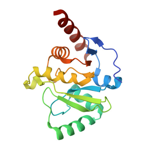
Viral macrodomains: a structural and evolutionary assessment of the pharmacological potential.

Rack, J.G.M.Zorzini, V.Zhu, Z.Schuller, M.Ahel, D.Ahel, I.

(2020) Open Biol 10: 200237-200237

  • DOI: https://doi.org/10.1098/rsob.200237
  • Primary Citation Related Structures: 
    6Z5T, 6Z6I, 6Z72

  • PubMed Abstract: 

    Viral macrodomains possess the ability to counteract host ADP-ribosylation, a post-translational modification implicated in the creation of an antiviral environment via immune response regulation. This brought them into focus as promising therapeutic targets, albeit the close homology to some of the human macrodomains raised concerns regarding potential cross-reactivity and adverse effects for the host. Here, we evaluate the structure and function of the macrodomain of SARS-CoV-2, the causative agent of COVID-19. We show that it can antagonize ADP-ribosylation by PARP14, a cellular (ADP-ribosyl)transferase necessary for the restriction of coronaviral infections. Furthermore, our structural studies together with ligand modelling revealed the structural basis for poly(ADP-ribose) binding and hydrolysis, an emerging new aspect of viral macrodomain biology. These new insights were used in an extensive evolutionary analysis aimed at evaluating the druggability of viral macrodomains not only from the Coronaviridae but also Togaviridae and Iridoviridae genera (causing diseases such as Chikungunya and infectious spleen and kidney necrosis virus disease, respectively). We found that they contain conserved features, distinct from their human counterparts, which may be exploited during drug design.


  • Organizational Affiliation
    • Sir William Dunn School of Pathology, University of Oxford, South Parks Road, Oxford OX1 3RE, UK.

Macromolecule Content 

  • Total Structure Weight: 80.85 kDa 
  • Atom Count: 6,354 
  • Modeled Residue Count: 692 
  • Deposited Residue Count: 704 
  • Unique protein chains: 1

Macromolecules

Find similar proteins by:|  3D Structure
Entity ID: 1
MoleculeChains  Sequence LengthOrganismDetailsImage
Replicase polyprotein 1ab
A, B, C, D
176Severe acute respiratory syndrome coronavirus 2Mutation(s): 0 
Gene Names: rep1a-1b
EC: 3.4.19.12 (PDB Primary Data), 3.4.22 (PDB Primary Data), 3.4.22.69 (PDB Primary Data), 2.7.7.48 (PDB Primary Data), 3.6.4.12 (PDB Primary Data), 3.6.4.13 (PDB Primary Data), 3.1.13 (PDB Primary Data), 3.1 (PDB Primary Data), 2.1.1 (PDB Primary Data)
UniProt
Find proteins for P0DTD1 (Severe acute respiratory syndrome coronavirus 2)
Explore P0DTD1 
Go to UniProtKB:  P0DTD1
Entity Groups
Sequence Clusters30% Identity50% Identity70% Identity90% Identity95% Identity100% Identity
UniProt GroupP0DTD1
Sequence Annotations
Expand
Reference Sequence

Small Molecules

Ligands 5 Unique
IDChains Name / Formula / InChI Key2D Diagram3D Interactions
A1R
(Subject of Investigation/LOI)

Query on A1R


Download:Ideal Coordinates CCD File
CA [auth D]
E [auth A]
J [auth A]
M [auth B]
R [auth C]
CA [auth D],
E [auth A],
J [auth A],
M [auth B],
R [auth C],
X [auth C]
5'-O-[(S)-{[(S)-{[(2R,3R,4S)-3,4-DIHYDROXYPYRROLIDIN-2-YL]METHOXY}(HYDROXY)PHOSPHORYL]OXY}(HYDROXY)PHOSPHORYL]ADENOSINE
C15 H24 N6 O12 P2
NDQDTBCXPOIQGT-UHNJQBFTSA-N
MPO

Query on MPO


Download:Ideal Coordinates CCD File
O [auth B]3[N-MORPHOLINO]PROPANE SULFONIC ACID
C7 H15 N O4 S
DVLFYONBTKHTER-UHFFFAOYSA-N
GOL

Query on GOL


Download:Ideal Coordinates CCD File
DA [auth D],
H [auth A],
N [auth B],
S [auth C],
W [auth C]
GLYCEROL
C3 H8 O3
PEDCQBHIVMGVHV-UHFFFAOYSA-N
EDO

Query on EDO


Download:Ideal Coordinates CCD File
EA [auth D]
F [auth A]
FA [auth D]
G [auth A]
GA [auth D]
EA [auth D],
F [auth A],
FA [auth D],
G [auth A],
GA [auth D],
HA [auth D],
I [auth A],
IA [auth D],
JA [auth D],
P [auth B],
T [auth C],
U [auth C],
V [auth C]
1,2-ETHANEDIOL
C2 H6 O2
LYCAIKOWRPUZTN-UHFFFAOYSA-N
NA

Query on NA


Download:Ideal Coordinates CCD File
AA [auth C]
BA [auth C]
K [auth A]
KA [auth D]
L [auth A]
AA [auth C],
BA [auth C],
K [auth A],
KA [auth D],
L [auth A],
LA [auth D],
Q [auth B],
Y [auth C],
Z [auth C]
SODIUM ION
Na
FKNQFGJONOIPTF-UHFFFAOYSA-N

Experimental Data & Validation

Experimental Data

  • Method: X-RAY DIFFRACTION
  • Resolution: 2.00 Å
  • R-Value Free:  0.279 (Depositor), 0.283 (DCC) 
  • R-Value Work:  0.229 (Depositor), 0.239 (DCC) 
  • R-Value Observed: 0.232 (Depositor) 
Space Group: P 1 21 1
Unit Cell:
Length ( Å )Angle ( ˚ )
a = 59.816α = 90
b = 83.096β = 94.51
c = 84.363γ = 90
Software Package:
Software NamePurpose
PHENIXrefinement
Aimlessdata scaling
PDB_EXTRACTdata extraction
xia2data reduction
PHASERphasing

Structure Validation

View Full Validation Report


Ligand Structure Quality Assessment 


Entry History 

& Funding Information

Deposition Data


Funding OrganizationLocationGrant Number
Wellcome TrustUnited Kingdom101794 and 210634

Revision History  (Full details and data files)

  • Version 1.0: 2020-12-02
    Type: Initial release
  • Version 1.1: 2024-01-24
    Changes: Data collection, Database references, Refinement description