


Crystal structure of haspin (GSG2) in complex with macrocycle ODS2003791
Chaikuad, A., Benderitter, P., Hoflack, J., Denis, A., Knapp, S., Structural Genomics Consortium (SGC)To be published.
Experimental Data Snapshot
Starting Model: experimental
View more details
Entity ID: 1 | |||||
|---|---|---|---|---|---|
| Molecule | Chains | Sequence Length | Organism | Details | Image |
| Serine/threonine-protein kinase haspin | 357 | Homo sapiens | Mutation(s): 0 Gene Names: HASPIN, GSG2 EC: 2.7.11.1 | 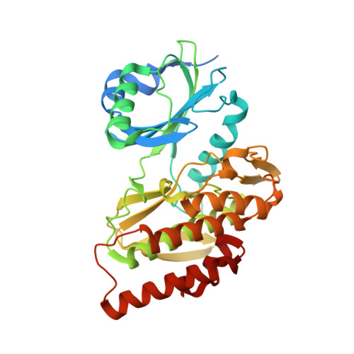 | |
UniProt & NIH Common Fund Data Resources | |||||
GTEx: ENSG00000177602 | |||||
Entity Groups | |||||
| Sequence Clusters | 30% Identity50% Identity70% Identity90% Identity95% Identity100% Identity | ||||
| UniProt Group | Q8TF76 | ||||
Sequence AnnotationsExpand | |||||
Reference Sequence | |||||
| Ligands 3 Unique | |||||
|---|---|---|---|---|---|
| ID | Chains | Name / Formula / InChI Key | 2D Diagram | 3D Interactions | |
| Q9B (Subject of Investigation/LOI) Download:Ideal Coordinates CCD File | B [auth A] | 8,14,18,19,22-pentazatetracyclo[13.5.2.12,6.018,21]tricosa-1(21),2,4,6(23),15(22),16,19-heptaen-7-one C18 H19 N5 O FZYFWWFZJSULHK-UHFFFAOYSA-N |  | ||
| MPD Download:Ideal Coordinates CCD File | D [auth A] | (4S)-2-METHYL-2,4-PENTANEDIOL C6 H14 O2 SVTBMSDMJJWYQN-YFKPBYRVSA-N |  | ||
| NA Download:Ideal Coordinates CCD File | C [auth A] | SODIUM ION Na FKNQFGJONOIPTF-UHFFFAOYSA-N |  | ||
| Length ( Å ) | Angle ( ˚ ) |
|---|---|
| a = 69.599 | α = 90 |
| b = 77.68 | β = 90 |
| c = 86.209 | γ = 90 |
| Software Name | Purpose |
|---|---|
| SCALA | data scaling |
| REFMAC | refinement |
| PDB_EXTRACT | data extraction |
| iMOSFLM | data reduction |
| PHASER | phasing |


