


Crystal structure of CLK1 in complex with macrocycle ODS2004070
Chaikuad, A., Benderitter, P., Hoflack, J., Denis, A., Knapp, S., Structural Genomics Consortium (SGC)To be published.
Experimental Data Snapshot
Starting Model: experimental
View more details
Entity ID: 1 | |||||
|---|---|---|---|---|---|
| Molecule | Chains | Sequence Length | Organism | Details | Image |
| Dual specificity protein kinase CLK1 | 339 | Homo sapiens | Mutation(s): 1 Gene Names: CLK1, CLK EC: 2.7.12.1 | 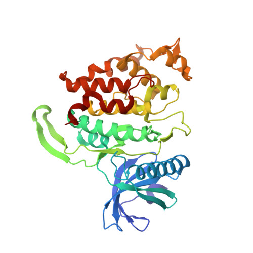 | |
UniProt & NIH Common Fund Data Resources | |||||
PHAROS: P49759 GTEx: ENSG00000013441 | |||||
Entity Groups | |||||
| Sequence Clusters | 30% Identity50% Identity70% Identity90% Identity95% Identity100% Identity | ||||
| UniProt Group | P49759 | ||||
Sequence AnnotationsExpand | |||||
Reference Sequence | |||||
| Ligands 3 Unique | |||||
|---|---|---|---|---|---|
| ID | Chains | Name / Formula / InChI Key | 2D Diagram | 3D Interactions | |
| PQ5 (Subject of Investigation/LOI) Download:Ideal Coordinates CCD File | H [auth A], I [auth B], K [auth C] | 7,10-Dioxa-13,17,18,21-tetrazatetracyclo[12.5.2.12,6.017,20]docosa-1(20),2(22),3,5,14(21),15,18-heptaene-5-carboxylic acid C17 H16 N4 O4 ZZVWSUCYWBUTCA-UHFFFAOYSA-N |  | ||
| PO4 Download:Ideal Coordinates CCD File | D [auth A], E [auth A], F [auth A], J [auth C] | PHOSPHATE ION O4 P NBIIXXVUZAFLBC-UHFFFAOYSA-K |  | ||
| GOL Download:Ideal Coordinates CCD File | G [auth A] | GLYCEROL C3 H8 O3 PEDCQBHIVMGVHV-UHFFFAOYSA-N |  | ||
| Length ( Å ) | Angle ( ˚ ) |
|---|---|
| a = 56.29 | α = 90 |
| b = 116.67 | β = 98.59 |
| c = 91.291 | γ = 90 |
| Software Name | Purpose |
|---|---|
| SCALA | data scaling |
| REFMAC | refinement |
| PDB_EXTRACT | data extraction |
| iMOSFLM | data reduction |
| PHASER | phasing |


