Insight into the RssB-Mediated Recognition and Delivery of sigma s to the AAA+ Protease, ClpXP.
Micevski, D., Zeth, K., Mulhern, T.D., Schuenemann, V.J., Zammit, J.E., Truscott, K.N., Dougan, D.A.(2020) Biomolecules 10
- PubMed: 32316259 Search on PubMedSearch on PubMed Central
- DOI: https://doi.org/10.3390/biom10040615
- Primary Citation Related Structures:
6Z4C, 6Z4E - PubMed Abstract:
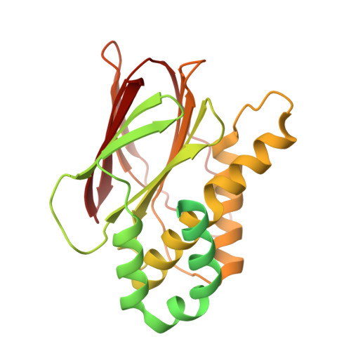
In Escherichia coli , SigmaS (σ S ) is the master regulator of the general stress response. The cellular levels of σ S are controlled by transcription, translation and protein stability. The turnover of σ S , by the AAA+ protease (ClpXP), is tightly regulated by a dedicated adaptor protein, termed RssB (Regulator of Sigma S protein B)-which is an atypical member of the response regulator (RR) family. Currently however, the molecular mechanism of σ S recognition and delivery by RssB is only poorly understood. Here we describe the crystal structures of both RssB domains (RssB N and RssB C ) and the SAXS analysis of full-length RssB (both free and in complex with σ S ). Together with our biochemical analysis we propose a model for the recognition and delivery of σ S by this essential adaptor protein. Similar to most bacterial RRs, the N-terminal domain of RssB (RssB N ) comprises a typical mixed (βα) 5 -fold. Although phosphorylation of RssB N (at Asp58) is essential for high affinity binding of σ S , much of the direct binding to σ S occurs via the C-terminal effector domain of RssB (RssB C ). In contrast to most RRs the effector domain of RssB forms a β-sandwich fold composed of two sheets surrounded by α-helical protrusions and as such, shares structural homology with serine/threonine phosphatases that exhibit a PPM/PP2C fold. Our biochemical data demonstrate that this domain plays a key role in both substrate interaction and docking to the zinc binding domain (ZBD) of ClpX. We propose that RssB docking to the ZBD of ClpX overlaps with the docking site of another regulator of RssB, the anti-adaptor IraD. Hence, we speculate that docking to ClpX may trigger release of its substrate through activation of a "closed" state (as seen in the RssB-IraD complex), thereby coupling adaptor docking (to ClpX) with substrate release. This competitive docking to RssB would prevent futile interaction of ClpX with the IraD-RssB complex (which lacks a substrate). Finally, substrate recognition by RssB appears to be regulated by a key residue (Arg117) within the α5 helix of the N-terminal domain. Importantly, this residue is not directly involved in σ S interaction, as σ S binding to the R117A mutant can be restored by phosphorylation. Likewise, R117A retains the ability to interact with and activate ClpX for degradation of σ S , both in the presence and absence of acetyl phosphate. Therefore, we propose that this region of RssB (the α5 helix) plays a critical role in driving interaction with σ S at a distal site.
- Department of Biochemistry and Genetics, La Trobe Institute for Molecular Science, La Trobe University, Melbourne 3086, Victoria, Australia.
Organizational Affiliation: