6Z3Z | pdb_00006z3z

CryoEM structure of horse sodium/proton exchanger NHE9 without C-terminal regulatory domain in an inward-facing conformation


Experimental Data Snapshot

  • Method: ELECTRON MICROSCOPY
  • Resolution: 3.19 Å
  • Aggregation State: PARTICLE 
  • Reconstruction Method: SINGLE PARTICLE 

wwPDB Validation 3D Report Full Report

Validation slider image for 6Z3Z

This is version 1.6 of the entry. See complete history

Literature

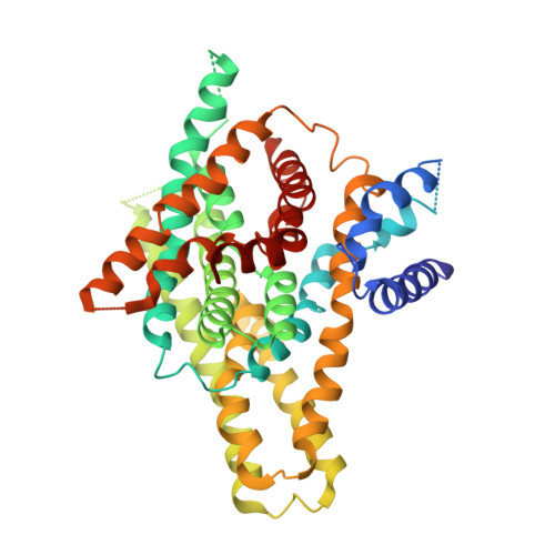
Structure and elevator mechanism of the mammalian sodium/proton exchanger NHE9.

Winklemann, I.Matsuoka, R.Meier, P.F.Shutin, D.Zhang, C.Orellana, L.Sexton, R.Landreh, M.Robinson, C.V.Beckstein, O.Drew, D.

(2020) EMBO J 39: e105908-e105908

  • DOI: https://doi.org/10.15252/embj.2020105908
  • Primary Citation Related Structures: 
    6Z3Y, 6Z3Z

  • PubMed Abstract: 

    Na + /H + exchangers (NHEs) are ancient membrane-bound nanomachines that work to regulate intracellular pH, sodium levels and cell volume. NHE activities contribute to the control of the cell cycle, cell proliferation, cell migration and vesicle trafficking. NHE dysfunction has been linked to many diseases, and they are targets of pharmaceutical drugs. Despite their fundamental importance to cell homeostasis and human physiology, structural information for the mammalian NHE was lacking. Here, we report the cryogenic electron microscopy structure of NHE isoform 9 (SLC9A9) from Equus caballus at 3.2 Å resolution, an endosomal isoform highly expressed in the brain and associated with autism spectrum (ASD) and attention deficit hyperactivity (ADHD) disorders. Despite low sequence identity, the NHE9 architecture and ion-binding site are remarkably similar to distantly related bacterial Na + /H +  antiporters with 13 transmembrane segments. Collectively, we reveal the conserved architecture of the NHE ion-binding site, their elevator-like structural transitions, the functional implications of autism disease mutations and the role of phosphoinositide lipids to promote homodimerization that, together, have important physiological ramifications.


  • Organizational Affiliation
    • Department of Biochemistry and Biophysics, Stockholm University, Stockholm, Sweden.

Macromolecule Content 

  • Total Structure Weight: 105.14 kDa 
  • Atom Count: 6,115 
  • Modeled Residue Count: 769 
  • Deposited Residue Count: 938 
  • Unique protein chains: 1

Macromolecules

Find similar proteins by:|  3D Structure
Entity ID: 1
MoleculeChains  Sequence LengthOrganismDetailsImage
Sodium/hydrogen exchangerA [auth B],
B [auth A]
469Equus caballusMutation(s): 0 
Gene Names: SLC9A9
Membrane Entity: Yes 
UniProt
Find proteins for F7B113 (Equus caballus)
Explore F7B113 
Go to UniProtKB:  F7B113
Entity Groups
Sequence Clusters30% Identity50% Identity70% Identity90% Identity95% Identity100% Identity
UniProt GroupF7B113
Sequence Annotations
Expand
Reference Sequence

Experimental Data & Validation

Experimental Data

  • Method: ELECTRON MICROSCOPY
  • Resolution: 3.19 Å
  • Aggregation State: PARTICLE 
  • Reconstruction Method: SINGLE PARTICLE 
EM Software:
TaskSoftware PackageVersion
MODEL REFINEMENTPHENIX
RECONSTRUCTIONRELION3.0

Structure Validation

View Full Validation Report


Entry History 

Deposition Data

Revision History  (Full details and data files)

  • Version 1.0: 2020-11-04
    Type: Initial release
  • Version 1.1: 2020-11-11
    Changes: Database references
  • Version 1.2: 2021-01-13
    Changes: Structure summary
  • Version 1.3: 2021-03-03
    Changes: Database references
  • Version 1.4: 2021-03-17
    Changes: Database references
  • Version 1.5: 2024-05-22
    Changes: Data collection, Database references
  • Version 1.6: 2025-07-02
    Changes: Data collection, Structure summary