Insights into the mechanisms of light-oxygen-voltage domain color tuning from a set of high-resolution X-ray structures.
Remeeva, A., Nazarenko, V.V., Kovalev, K., Goncharov, I.M., Yudenko, A., Astashkin, R., Gordeliy, V., Gushchin, I.(2021) Proteins
- PubMed: 33774867 Search on PubMed
- DOI: https://doi.org/10.1002/prot.26078
- Primary Citation Related Structures:
6YWG, 6YWH, 6YWI, 6YWQ, 6YWR, 6YXC - PubMed Abstract:
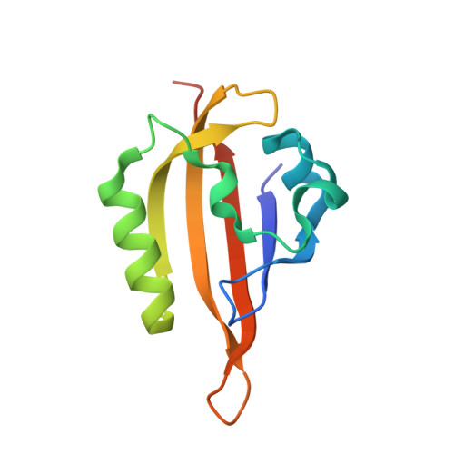
Light-oxygen-voltage (LOV) domains are widespread photosensory modules that can be used in fluorescence microscopy, optogenetics and controlled production of reactive oxygen species. All of the currently known LOV domains have absorption maxima in the range of ~440 to ~450 nm, and it is not clear whether they can be shifted significantly using mutations. Here, we have generated a panel of LOV domain variants by mutating the key chromophore-proximal glutamine aminoacid of a thermostable flavin based fluorescent protein CagFbFP (Gln148) to asparagine, aspartate, glutamate, histidine, lysine and arginine. Absorption spectra of all of the mutants are blue-shifted, with the maximal shift of 8 nm observed for the Q148H variant. While CagFbFP and its Q148N/D/E variants are not sensitive to pH, Q148H/K/R reveal a moderate red shift induced byacidic pH. To gain further insight, we determined high resolution crystal structures of all of the mutants studied at the resolutions from 1.07 Å for Q148D to 1.63 Å for Q148R. Whereas in some of the variants, the aminoacid 148 remains in the vicinity of the flavin, in Q148K, Q148R and partially Q148D, the C-terminus of the protein unlatches and the side chain of the residue 148 is reoriented away from the chromophore. Our results explain the absence of color shifts from replacing Gln148 with charged aminoacids and pave the way for rational design of color-shifted flavin based fluorescent proteins.
- Research Center for Molecular Mechanisms of Aging and Age-Related Diseases, Moscow Institute of Physics and Technology, Dolgoprudny, Russia.
Organizational Affiliation: