6YWD

De novo designed protein 4H_01 in complex with Mota antibody


Experimental Data Snapshot

  • Method: X-RAY DIFFRACTION
  • Resolution: 3.20 Å
  • R-Value Free: 0.247 
  • R-Value Work: 0.206 
  • R-Value Observed: 0.210 

wwPDB Validation   3D Report Full Report


This is version 1.3 of the entry. See complete history


Literature

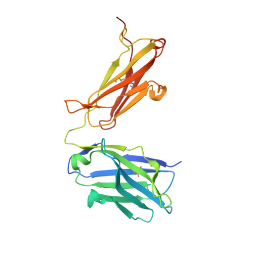
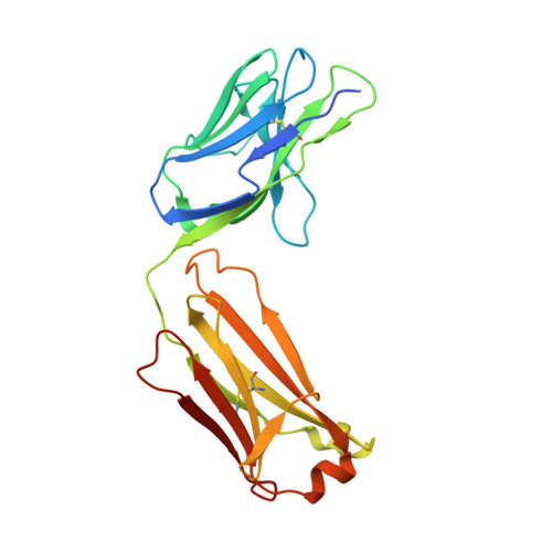
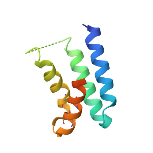
Bottom-up de novo design of functional proteins with complex structural features.

Yang, C.Sesterhenn, F.Bonet, J.van Aalen, E.A.Scheller, L.Abriata, L.A.Cramer, J.T.Wen, X.Rosset, S.Georgeon, S.Jardetzky, T.Krey, T.Fussenegger, M.Merkx, M.Correia, B.E.

(2021) Nat Chem Biol 17: 492-500

  • DOI: https://doi.org/10.1038/s41589-020-00699-x
  • Primary Citation of Related Structures:  
    6YWC, 6YWD

  • PubMed Abstract: 

    De novo protein design has enabled the creation of new protein structures. However, the design of functional proteins has proved challenging, in part due to the difficulty of transplanting structurally complex functional sites to available protein structures. Here, we used a bottom-up approach to build de novo proteins tailored to accommodate structurally complex functional motifs. We applied the bottom-up strategy to successfully design five folds for four distinct binding motifs, including a bifunctionalized protein with two motifs. Crystal structures confirmed the atomic-level accuracy of the computational designs. These de novo proteins were functional as components of biosensors to monitor antibody responses and as orthogonal ligands to modulate synthetic signaling receptors in engineered mammalian cells. Our work demonstrates the potential of bottom-up approaches to accommodate complex structural motifs, which will be essential to endow de novo proteins with elaborate biochemical functions, such as molecular recognition or catalysis.


  • Organizational Affiliation

    Institute of Bioengineering, École Polytechnique Fédérale de Lausanne (EPFL), Lausanne, Switzerland.


Macromolecules
Find similar proteins by:  (by identity cutoff)  |  3D Structure
Entity ID: 1
MoleculeChains Sequence LengthOrganismDetailsImage
Antibody Mota, Heavy Chain233Homo sapiensMutation(s): 0 
Entity Groups  
Sequence Clusters30% Identity50% Identity70% Identity90% Identity95% Identity100% Identity
Sequence Annotations
Expand
  • Reference Sequence
Find similar proteins by:  (by identity cutoff)  |  3D Structure
Entity ID: 2
MoleculeChains Sequence LengthOrganismDetailsImage
Antibody Mota, Light Chain221Homo sapiensMutation(s): 0 
Entity Groups  
Sequence Clusters30% Identity50% Identity70% Identity90% Identity95% Identity100% Identity
Sequence Annotations
Expand
  • Reference Sequence
Find similar proteins by:  (by identity cutoff)  |  3D Structure
Entity ID: 3
MoleculeChains Sequence LengthOrganismDetailsImage
De novo designed protein 4H_0191synthetic constructMutation(s): 0 
Entity Groups  
Sequence Clusters30% Identity50% Identity70% Identity90% Identity95% Identity100% Identity
Sequence Annotations
Expand
  • Reference Sequence
Experimental Data & Validation

Experimental Data

  • Method: X-RAY DIFFRACTION
  • Resolution: 3.20 Å
  • R-Value Free: 0.247 
  • R-Value Work: 0.206 
  • R-Value Observed: 0.210 
  • Space Group: P 31 2 1
Unit Cell:
Length ( Å )Angle ( ˚ )
a = 143.71α = 90
b = 143.71β = 90
c = 90.223γ = 120
Software Package:
Software NamePurpose
PHENIXrefinement
PHENIXrefinement
XDSdata reduction
XDSdata scaling
PHENIXphasing

Structure Validation

View Full Validation Report


Entry History & Funding Information

Deposition Data


Funding OrganizationLocationGrant Number
Swiss National Science FoundationSwitzerland2015/333
European CommissionEuropean Union716058
Swiss National Science FoundationSwitzerland310030-163139

Revision History  (Full details and data files)

  • Version 1.0: 2020-10-07
    Type: Initial release
  • Version 1.1: 2021-01-20
    Changes: Database references
  • Version 1.2: 2021-04-07
    Changes: Database references
  • Version 1.3: 2024-01-24
    Changes: Data collection, Database references, Refinement description