6YV4 | pdb_00006yv4

STRUCTURE OF THE WNT DEACYLASE NOTUM IN COMPLEX WITH A PYRROLE-3-CARBOXYLIC ACID FRAGMENT 686


Experimental Data Snapshot

  • Method: X-RAY DIFFRACTION
  • Resolution: 2.00 Å
  • R-Value Free: 
    0.233 (Depositor), 0.235 (DCC) 
  • R-Value Work: 
    0.181 (Depositor), 0.184 (DCC) 
  • R-Value Observed: 
    0.184 (Depositor) 

Starting Model: experimental
View more details

wwPDB Validation 3D Report Full Report

Validation slider image for 6YV4

Ligand Structure Quality Assessment 


This is version 1.6 of the entry. See complete history

Literature

Screening of a Custom-Designed Acid Fragment Library Identifies 1-Phenylpyrroles and 1-Phenylpyrrolidines as Inhibitors of Notum Carboxylesterase Activity.

Mahy, W.Patel, M.Steadman, D.Woodward, H.L.Atkinson, B.N.Svensson, F.Willis, N.J.Flint, A.Papatheodorou, D.Zhao, Y.Vecchia, L.Ruza, R.R.Hillier, J.Frew, S.Monaghan, A.Costa, A.Bictash, M.Walter, M.W.Jones, E.Y.Fish, P.V.

(2020) J Med Chem 63: 9464-9483

  • DOI: https://doi.org/10.1021/acs.jmedchem.0c00660
  • Primary Citation Related Structures: 
    6YSK, 6YUW, 6YUY, 6YV0, 6YV2, 6YV4, 6YXI

  • PubMed Abstract: 

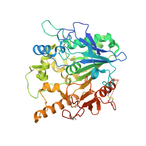
    The Wnt family of proteins are secreted signaling proteins that play key roles in regulating cellular functions. Recently, carboxylesterase Notum was shown to act as a negative regulator of Wnt signaling by mediating the removal of an essential palmitoleate. Here we disclose two new chemical scaffolds that inhibit Notum enzymatic activity. Our approach was to create a fragment library of 250 acids for screening against Notum in a biochemical assay followed by structure determination by X-ray crystallography. Twenty fragments were identified as hits for Notum inhibition, and 14 of these fragments were shown to bind in the palmitoleate pocket of Notum. Optimization of 1-phenylpyrrole 20 , guided by structure-based drug design, identified 20z as the most potent compound from this series. Similarly, the optimization of 1-phenylpyrrolidine 8 gave acid 26 . This work demonstrates that inhibition of Notum activity can be achieved by small, drug-like molecules possessing favorable in vitro ADME profiles.


  • Organizational Affiliation
    • Alzheimer's Research UK UCL Drug Discovery Institute, University College London, Cruciform Building, Gower Street, London WC1E 6BT, United Kingdom.

Macromolecule Content 

  • Total Structure Weight: 44.47 kDa 
  • Atom Count: 3,103 
  • Modeled Residue Count: 359 
  • Deposited Residue Count: 383 
  • Unique protein chains: 1

Macromolecules

Find similar proteins by:|  3D Structure
Entity ID: 1
MoleculeChains  Sequence LengthOrganismDetailsImage
Palmitoleoyl-protein carboxylesterase NOTUM383Homo sapiensMutation(s): 1 
Gene Names: NOTUMOK/SW-CL.30
EC: 3.1.1.98
UniProt & NIH Common Fund Data Resources
Find proteins for Q6P988 (Homo sapiens)
Explore Q6P988 
Go to UniProtKB:  Q6P988
PHAROS:  Q6P988
GTEx:  ENSG00000185269 
Entity Groups
Sequence Clusters30% Identity50% Identity70% Identity90% Identity95% Identity100% Identity
UniProt GroupQ6P988
Glycosylation
Glycosylation Sites: 1Go to GlyGen: Q6P988-1
Sequence Annotations
Expand
Reference Sequence

Small Molecules

Ligands 4 Unique
IDChains Name / Formula / InChI Key2D Diagram3D Interactions
NAG

Query on NAG


Download:Ideal Coordinates CCD File
C [auth A]2-acetamido-2-deoxy-beta-D-glucopyranose
C8 H15 N O6
OVRNDRQMDRJTHS-FMDGEEDCSA-N
PQK
(Subject of Investigation/LOI)

Query on PQK


Download:Ideal Coordinates CCD File
B [auth A]1-cyclopropyl-2,5-dimethyl-pyrrole-3-carboxylic acid
C10 H13 N O2
VOOZJPFNBFNPEK-UHFFFAOYSA-N
SO4

Query on SO4


Download:Ideal Coordinates CCD File
D [auth A],
J [auth A]
SULFATE ION
O4 S
QAOWNCQODCNURD-UHFFFAOYSA-L
EDO

Query on EDO


Download:Ideal Coordinates CCD File
E [auth A],
F [auth A],
G [auth A],
H [auth A],
I [auth A]
1,2-ETHANEDIOL
C2 H6 O2
LYCAIKOWRPUZTN-UHFFFAOYSA-N
Binding Affinity Annotations 
IDSourceBinding Affinity
PQK BindingDB:  6YV4 IC50: 6300 (nM) from 1 assay(s)

Experimental Data & Validation

Experimental Data

  • Method: X-RAY DIFFRACTION
  • Resolution: 2.00 Å
  • R-Value Free:  0.233 (Depositor), 0.235 (DCC) 
  • R-Value Work:  0.181 (Depositor), 0.184 (DCC) 
  • R-Value Observed: 0.184 (Depositor) 
Space Group: P 21 21 21
Unit Cell:
Length ( Å )Angle ( ˚ )
a = 59.24α = 90
b = 73.46β = 90
c = 79.38γ = 90
Software Package:
Software NamePurpose
PHENIXrefinement
xia2data reduction
DIALSdata scaling
PHASERphasing

Structure Validation

View Full Validation Report


Ligand Structure Quality Assessment 


Entry History 

& Funding Information

Deposition Data


Funding OrganizationLocationGrant Number
Cancer Research UKUnited KingdomC375/A17721

Revision History  (Full details and data files)

  • Version 1.0: 2020-05-06
    Type: Initial release
  • Version 1.1: 2020-06-03
    Changes: Structure summary
  • Version 1.2: 2020-07-29
    Type: Remediation
    Reason: Carbohydrate remediation
    Changes: Data collection, Derived calculations, Structure summary
  • Version 1.3: 2020-08-26
    Changes: Database references, Structure summary
  • Version 1.4: 2020-09-23
    Changes: Database references
  • Version 1.5: 2024-01-24
    Changes: Data collection, Database references, Refinement description
  • Version 1.6: 2024-10-09
    Changes: Structure summary