6YQ1 | pdb_00006yq1

FOCAL ADHESION KINASE CATALYTIC DOMAIN IN COMPLEX WITH N-Methyl-N-(3-{[2-(2-oxo-1,2,3,4-tetrahydro-quinolin-6-ylamino)-5-trifluoromethyl-pyrimidin-4-ylamino]-methyl}-pyridin-2-yl)-methanesulfonamide


Experimental Data Snapshot

  • Method: X-RAY DIFFRACTION
  • Resolution: 1.78 Å
  • R-Value Free: 
    0.214 (Depositor) 
  • R-Value Work: 
    0.183 (Depositor) 
  • R-Value Observed: 
    0.184 (Depositor) 

Starting Model: experimental
View more details

wwPDB Validation 3D Report Full Report

Validation slider image for 6YQ1

Ligand Structure Quality Assessment 


This is version 1.3 of the entry. See complete history

Literature

Structure-kinetic relationship reveals the mechanism of selectivity of FAK inhibitors over PYK2.

Berger, B.T.Amaral, M.Kokh, D.B.Nunes-Alves, A.Musil, D.Heinrich, T.Schroder, M.Neil, R.Wang, J.Navratilova, I.Bomke, J.Elkins, J.M.Muller, S.Frech, M.Wade, R.C.Knapp, S.

(2021) Cell Chem Biol 28: 686

  • DOI: https://doi.org/10.1016/j.chembiol.2021.01.003
  • Primary Citation Related Structures: 
    6YOJ, 6YQ1, 6YR9, 6YT6, 6YVS, 6YVY, 6YXV

  • PubMed Abstract: 

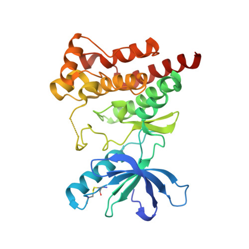
    There is increasing evidence of a significant correlation between prolonged drug-target residence time and increased drug efficacy. Here, we report a structural rationale for kinetic selectivity between two closely related kinases: focal adhesion kinase (FAK) and proline-rich tyrosine kinase 2 (PYK2). We found that slowly dissociating FAK inhibitors induce helical structure at the DFG motif of FAK but not PYK2. Binding kinetic data, high-resolution structures and mutagenesis data support the role of hydrophobic interactions of inhibitors with the DFG-helical region, providing a structural rationale for slow dissociation rates from FAK and kinetic selectivity over PYK2. Our experimental data correlate well with computed relative residence times from molecular simulations, supporting a feasible strategy for rationally optimizing ligand residence times. We suggest that the interplay between the protein structural mobility and ligand-induced effects is a key regulator of the kinetic selectivity of inhibitors of FAK versus PYK2.


  • Organizational Affiliation
    • Structural Genomics Consortium, Goethe University Frankfurt, Buchmann Institute for Molecular Life Sciences, Max-von-Laue-Straße 15, 60438 Frankfurt am Main, Germany; Institute of Pharmaceutical Chemistry, Goethe University Frankfurt, Buchmann Institute for Molecular Life Sciences, Max-von-Laue-Straße 9, 60438 Frankfurt am Main, Germany.

Macromolecule Content 

  • Total Structure Weight: 131.42 kDa 
  • Atom Count: 9,732 
  • Modeled Residue Count: 1,065 
  • Deposited Residue Count: 1,128 
  • Unique protein chains: 1

Macromolecules

Find similar proteins by:|  3D Structure
Entity ID: 1
MoleculeChains  Sequence LengthOrganismDetailsImage
Focal adhesion kinase 1
A, B, C, D
282Homo sapiensMutation(s): 0 
Gene Names: PTK2FAKFAK1
EC: 2.7.10.2
UniProt & NIH Common Fund Data Resources
Find proteins for Q05397 (Homo sapiens)
Explore Q05397 
Go to UniProtKB:  Q05397
PHAROS:  Q05397
GTEx:  ENSG00000169398 
Entity Groups
Sequence Clusters30% Identity50% Identity70% Identity90% Identity95% Identity100% Identity
UniProt GroupQ05397
Sequence Annotations
Expand
Reference Sequence

Small Molecules

Ligands 3 Unique
IDChains Name / Formula / InChI Key2D Diagram3D Interactions
P7N
(Subject of Investigation/LOI)

Query on P7N


Download:Ideal Coordinates CCD File
E [auth A],
H [auth B],
I [auth C],
J [auth D]
~{N}-methyl-~{N}-[3-[[[2-[(2-oxidanylidene-3,4-dihydro-1~{H}-quinolin-6-yl)amino]-5-(trifluoromethyl)pyrimidin-4-yl]amino]methyl]pyridin-2-yl]methanesulfonamide
C22 H22 F3 N7 O3 S
MUVVMGFRHXQCRG-UHFFFAOYSA-N
SO4

Query on SO4


Download:Ideal Coordinates CCD File
G [auth A]SULFATE ION
O4 S
QAOWNCQODCNURD-UHFFFAOYSA-L
NA

Query on NA


Download:Ideal Coordinates CCD File
F [auth A]SODIUM ION
Na
FKNQFGJONOIPTF-UHFFFAOYSA-N
Binding Affinity Annotations 
IDSourceBinding Affinity
P7N BindingDB:  6YQ1 Ki: 3.2 (nM) from 1 assay(s)

Experimental Data & Validation

Experimental Data

  • Method: X-RAY DIFFRACTION
  • Resolution: 1.78 Å
  • R-Value Free:  0.214 (Depositor) 
  • R-Value Work:  0.183 (Depositor) 
  • R-Value Observed: 0.184 (Depositor) 
Space Group: C 1 2 1
Unit Cell:
Length ( Å )Angle ( ˚ )
a = 112.532α = 90
b = 75.749β = 102.37
c = 173.265γ = 90
Software Package:
Software NamePurpose
BUSTERrefinement
PDB_EXTRACTdata extraction
XDSdata reduction
autoPROCdata scaling
PHASERphasing

Structure Validation

View Full Validation Report


Ligand Structure Quality Assessment 


Entry History 

Deposition Data

Revision History  (Full details and data files)

  • Version 1.0: 2021-02-10
    Type: Initial release
  • Version 1.1: 2021-06-02
    Changes: Database references
  • Version 1.2: 2024-01-24
    Changes: Data collection, Database references, Refinement description
  • Version 1.3: 2024-11-13
    Changes: Structure summary