6YMZ | pdb_00006ymz

Structure of the CheB methylsterase from P. atrosepticum SCRI1043


Experimental Data Snapshot

  • Method: X-RAY DIFFRACTION
  • Resolution: 2.30 Å
  • R-Value Free: 
    0.226 (Depositor), 0.225 (DCC) 
  • R-Value Work: 
    0.192 (Depositor), 0.193 (DCC) 
  • R-Value Observed: 
    0.194 (Depositor) 

Starting Model: experimental
View more details

wwPDB Validation 3D Report Full Report

Validation slider image for 6YMZ

This is version 1.1 of the entry. See complete history

Literature

Evidence for Pentapeptide-Dependent and Independent CheB Methylesterases.

Velando, F.Gavira, J.A.Rico-Jimenez, M.Matilla, M.A.Krell, T.

(2020) Int J Mol Sci 21

  • DOI: https://doi.org/10.3390/ijms21228459
  • Primary Citation Related Structures: 
    6YMZ

  • PubMed Abstract: 

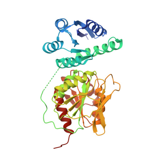
    Many bacteria possess multiple chemosensory pathways that are composed of homologous signaling proteins. These pathways appear to be functionally insulated from each other, but little information is available on the corresponding molecular basis. We report here a novel mechanism that contributes to pathway insulation. We show that, of the four CheB paralogs of Pseudomonas aeruginosa PAO1, only CheB 2 recognizes a pentapeptide at the C-terminal extension of the McpB (Aer2) chemoreceptor ( K D = 93 µM). McpB is the sole chemoreceptor that stimulates the Che2 pathway, and CheB 2 is the methylesterase of this pathway. Pectobacterium atrosepticum SCRI1043 has a single CheB, CheB_Pec, and 19 of its 36 chemoreceptors contain a C-terminal pentapeptide. The deletion of cheB_Pec abolished chemotaxis, but, surprisingly, none of the pentapeptides bound to CheB_Pec. To determine the corresponding structural basis, we solved the 3D structure of CheB_Pec. Its structure aligned well with that of the pentapeptide-dependent enzyme from Salmonella enterica . However, no electron density was observed in the CheB_Pec region corresponding to the pentapeptide-binding site in the Escherichia coli CheB. We hypothesize that this structural disorder is associated with the failure to bind pentapeptides. Combined data show that CheB methylesterases can be divided into pentapeptide-dependent and independent enzymes.


  • Organizational Affiliation
    • Department of Environmental Protection, Estación Experimental del Zaidín, Consejo Superior de Investigaciones Científicas, Prof. Albareda 1, 18008 Granada, Spain.

Macromolecule Content 

  • Total Structure Weight: 194.06 kDa 
  • Atom Count: 13,986 
  • Modeled Residue Count: 1,688 
  • Deposited Residue Count: 1,780 
  • Unique protein chains: 1

Macromolecules

Find similar proteins by:|  3D Structure
Entity ID: 1
MoleculeChains  Sequence LengthOrganismDetailsImage
Protein-glutamate methylesterase/protein-glutamine glutaminase
A, B, C, D, E
356Pectobacterium atrosepticum SCRI1043Mutation(s): 0 
Gene Names: cheBECA1693
EC: 3.1.1.61 (PDB Primary Data), 3.5.1.44 (PDB Primary Data)
UniProt
Find proteins for Q6D6I7 (Pectobacterium atrosepticum (strain SCRI 1043 / ATCC BAA-672))
Explore Q6D6I7 
Go to UniProtKB:  Q6D6I7
Entity Groups
Sequence Clusters30% Identity50% Identity70% Identity90% Identity95% Identity100% Identity
UniProt GroupQ6D6I7
Sequence Annotations
Expand
Reference Sequence

Small Molecules

Ligands 4 Unique
IDChains Name / Formula / InChI Key2D Diagram3D Interactions
SO4

Query on SO4


Download:Ideal Coordinates CCD File
T [auth E]SULFATE ION
O4 S
QAOWNCQODCNURD-UHFFFAOYSA-L
GOL

Query on GOL


Download:Ideal Coordinates CCD File
H [auth A]
J [auth A]
M [auth C]
P [auth C]
R [auth D]
H [auth A],
J [auth A],
M [auth C],
P [auth C],
R [auth D],
U [auth E],
V [auth E]
GLYCEROL
C3 H8 O3
PEDCQBHIVMGVHV-UHFFFAOYSA-N
ACT

Query on ACT


Download:Ideal Coordinates CCD File
F [auth A]ACETATE ION
C2 H3 O2
QTBSBXVTEAMEQO-UHFFFAOYSA-M
NA

Query on NA


Download:Ideal Coordinates CCD File
G [auth A]
I [auth A]
K [auth B]
L [auth B]
N [auth C]
G [auth A],
I [auth A],
K [auth B],
L [auth B],
N [auth C],
O [auth C],
Q [auth D],
S [auth E]
SODIUM ION
Na
FKNQFGJONOIPTF-UHFFFAOYSA-N

Experimental Data & Validation

Experimental Data

  • Method: X-RAY DIFFRACTION
  • Resolution: 2.30 Å
  • R-Value Free:  0.226 (Depositor), 0.225 (DCC) 
  • R-Value Work:  0.192 (Depositor), 0.193 (DCC) 
  • R-Value Observed: 0.194 (Depositor) 
Space Group: I 4
Unit Cell:
Length ( Å )Angle ( ˚ )
a = 148.741α = 90
b = 148.741β = 90
c = 206.34γ = 90
Software Package:
Software NamePurpose
PHASERphasing
PHENIXrefinement
MOSFLMdata reduction
Aimlessdata scaling
Cootmodel building

Structure Validation

View Full Validation Report


Entry History 

& Funding Information

Deposition Data


Funding OrganizationLocationGrant Number
Spanish Ministry of Science, Innovation, and UniversitiesSpainBIO2013-4297-P
Spanish Ministry of Science, Innovation, and UniversitiesSpainBIO2016-74875-P

Revision History  (Full details and data files)

  • Version 1.0: 2021-02-17
    Type: Initial release
  • Version 1.1: 2024-01-24
    Changes: Data collection, Database references, Refinement description