6YFA | pdb_00006yfa

Virus-like particle of bacteriophage AVE015


Experimental Data Snapshot

  • Method: X-RAY DIFFRACTION
  • Resolution: 3.30 Å
  • R-Value Free: 
    0.233 (Depositor), 0.233 (DCC) 
  • R-Value Work: 
    0.231 (Depositor), 0.231 (DCC) 
  • R-Value Observed: 
    0.231 (Depositor) 

Starting Model: experimental
View more details

wwPDB Validation 3D Report Full Report

Validation slider image for 6YFA

This is version 1.2 of the entry. See complete history

Literature

Three-dimensional structure of 22 uncultured ssRNA bacteriophages: Flexibility of the coat protein fold and variations in particle shapes.

Rumnieks, J.Lieknina, I.Kalnins, G.Sisovs, M.Akopjana, I.Bogans, J.Tars, K.

(2020) Sci Adv 6

  • DOI: https://doi.org/10.1126/sciadv.abc0023
  • Primary Citation Related Structures: 
    6YF7, 6YF9, 6YFA, 6YFB, 6YFC, 6YFD, 6YFE, 6YFF, 6YFG, 6YFH, 6YFI, 6YFJ, 6YFK, 6YFL, 6YFM, 6YFN, 6YFO, 6YFP, 6YFQ, 6YFR, 6YFS, 6YFT, 6YFU

  • PubMed Abstract: 

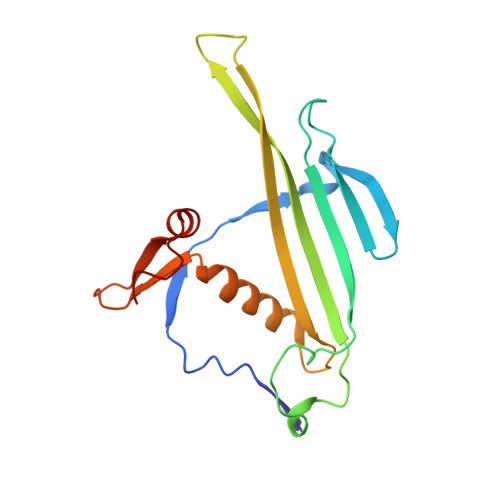
    The single-stranded RNA (ssRNA) bacteriophages are among the simplest known viruses with small genomes and exceptionally high mutation rates. The number of ssRNA phage isolates has remained very low, but recent metagenomic studies have uncovered an immense variety of distinct uncultured ssRNA phages. The coat proteins (CPs) in these genomes are particularly diverse, with notable variation in length and often no recognizable similarity to previously known viruses. We recombinantly expressed metagenome-derived ssRNA phage CPs to produce virus-like particles and determined the three-dimensional structure of 22 previously uncharacterized ssRNA phage capsids covering nine distinct CP types. The structures revealed substantial deviations from the previously known ssRNA phage CP fold, uncovered an unusual prolate particle shape, and revealed a previously unseen dsRNA binding mode. These data expand our knowledge of the evolution of viral structural proteins and are of relevance for applications such as ssRNA phage-based vaccine design.


  • Organizational Affiliation
    • Latvian Biomedical Research and Study Center, Rātsupītes 1, LV1067, Riga, Latvia.

Macromolecule Content 

  • Total Structure Weight: 1,121.65 kDa 
  • Atom Count: 78,980 
  • Modeled Residue Count: 10,020 
  • Deposited Residue Count: 10,020 
  • Unique protein chains: 1

Small Molecules

Ligands 1 Unique
IDChains Name / Formula / InChI Key2D Diagram3D Interactions
CA

Query on CA


Download:Ideal Coordinates CCD File
AC [auth CC]
BC [auth CF]
IB [auth AA]
JB [auth AD]
KB [auth AG]
AC [auth CC],
BC [auth CF],
IB [auth AA],
JB [auth AD],
KB [auth AG],
LB [auth AJ],
MB [auth AM],
NB [auth AP],
OB [auth AS],
PB [auth AV],
QB [auth AY],
RB [auth BB],
SB [auth BE],
TB [auth BH],
UB [auth BK],
VB [auth BN],
WB [auth BQ],
XB [auth BT],
YB [auth BW],
ZB [auth BZ]
CALCIUM ION
Ca
BHPQYMZQTOCNFJ-UHFFFAOYSA-N

Experimental Data & Validation

Experimental Data

  • Method: X-RAY DIFFRACTION
  • Resolution: 3.30 Å
  • R-Value Free:  0.233 (Depositor), 0.233 (DCC) 
  • R-Value Work:  0.231 (Depositor), 0.231 (DCC) 
  • R-Value Observed: 0.231 (Depositor) 
Space Group: I 2 3
Unit Cell:
Length ( Å )Angle ( ˚ )
a = 559.31α = 90
b = 559.31β = 90
c = 559.31γ = 90
Software Package:
Software NamePurpose
PHENIXrefinement
MOSFLMdata reduction
SCALAdata scaling
PDB_EXTRACTdata extraction
FFTphasing

Structure Validation

View Full Validation Report


Entry History 

& Funding Information

Deposition Data


Funding OrganizationLocationGrant Number
European Regional Development FundLatvia1.1.1.1/16/A/104

Revision History  (Full details and data files)

  • Version 1.0: 2020-09-02
    Type: Initial release
  • Version 1.1: 2020-12-16
    Changes: Database references
  • Version 1.2: 2024-01-24
    Changes: Data collection, Database references, Derived calculations, Refinement description