6YES | pdb_00006yes

Crystal structure of type I-D CRISPR-Cas nuclease Cas10d


Experimental Data Snapshot

  • Method: X-RAY DIFFRACTION
  • Resolution: 4.10 Å
  • R-Value Free: 
    0.370 (Depositor), 0.371 (DCC) 
  • R-Value Work: 
    0.343 (Depositor), 0.346 (DCC) 
  • R-Value Observed: 
    0.345 (Depositor) 

Starting Model: experimental
View more details

wwPDB Validation 3D Report Full Report

Validation slider image for 6YES

This is version 1.5 of the entry. See complete history

Literature

Structural basis for inhibition of an archaeal CRISPR-Cas type I-D large subunit by an anti-CRISPR protein.

Manav, M.C.Van, L.B.Lin, J.Fuglsang, A.Peng, X.Brodersen, D.E.

(2020) Nat Commun 11: 5993-5993

  • DOI: https://doi.org/10.1038/s41467-020-19847-x
  • Primary Citation Related Structures: 
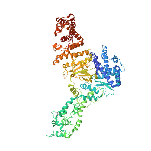
    6THH, 6YES

  • PubMed Abstract: 

    A hallmark of type I CRISPR-Cas systems is the presence of Cas3, which contains both the nuclease and helicase activities required for DNA cleavage during interference. In subtype I-D systems, however, the histidine-aspartate (HD) nuclease domain is encoded as part of a Cas10-like large effector complex subunit and the helicase activity in a separate Cas3' subunit, but the functional and mechanistic consequences of this organisation are not currently understood. Here we show that the Sulfolobus islandicus type I-D Cas10d large subunit exhibits an unusual domain architecture consisting of a Cas3-like HD nuclease domain fused to a degenerate polymerase fold and a C-terminal domain structurally similar to Cas11. Crystal structures of Cas10d both in isolation and bound to S. islandicus rod-shaped virus 3 AcrID1 reveal that the anti-CRISPR protein sequesters the large subunit in a non-functional state unable to form a cleavage-competent effector complex. The architecture of Cas10d suggests that the type I-D effector complex is similar to those found in type III CRISPR-Cas systems and that this feature is specifically exploited by phages for anti-CRISPR defence.


  • Organizational Affiliation
    • Department of Molecular Biology and Genetics, Aarhus University, Gustav Wieds Vej 10c, DK-8000, Aarhus C, Denmark.

Macromolecule Content 

  • Total Structure Weight: 195.11 kDa 
  • Atom Count: 12,534 
  • Modeled Residue Count: 1,567 
  • Deposited Residue Count: 1,710 
  • Unique protein chains: 1

Macromolecules

Find similar proteins by:|  3D Structure
Entity ID: 1
MoleculeChains  Sequence LengthOrganismDetailsImage
CRISPR-associated protein, CscA
A, B
855Saccharolobus islandicus LAL14/1Mutation(s): 0 
Gene Names: SiL_0609
UniProt
Find proteins for M9U4Y8 (Saccharolobus islandicus LAL14/1)
Explore M9U4Y8 
Go to UniProtKB:  M9U4Y8
Entity Groups
Sequence Clusters30% Identity50% Identity70% Identity90% Identity95% Identity100% Identity
UniProt GroupM9U4Y8
Sequence Annotations
Expand
Reference Sequence

Experimental Data & Validation

Experimental Data

  • Method: X-RAY DIFFRACTION
  • Resolution: 4.10 Å
  • R-Value Free:  0.370 (Depositor), 0.371 (DCC) 
  • R-Value Work:  0.343 (Depositor), 0.346 (DCC) 
  • R-Value Observed: 0.345 (Depositor) 
Space Group: P 43 21 2
Unit Cell:
Length ( Å )Angle ( ˚ )
a = 141.43α = 90
b = 141.43β = 90
c = 223.87γ = 90
Software Package:
Software NamePurpose
PHENIXrefinement
XDSdata reduction
XSCALEdata scaling
PHASERphasing
PDB_EXTRACTdata extraction

Structure Validation

View Full Validation Report


Entry History 

& Funding Information

Deposition Data


Funding OrganizationLocationGrant Number
LundbeckfondenDenmarkR-287-2018-1555
Novo Nordisk FoundationDenmarkNNF17OC0028072

Revision History  (Full details and data files)

  • Version 1.0: 2020-10-28
    Type: Initial release
  • Version 1.1: 2020-12-02
    Changes: Database references
  • Version 1.2: 2020-12-09
    Changes: Database references
  • Version 1.3: 2021-06-30
    Changes: Refinement description, Source and taxonomy, Structure summary
  • Version 1.4: 2024-01-24
    Changes: Data collection, Database references, Refinement description
  • Version 1.5: 2024-11-13
    Changes: Structure summary