6YCA

Crystal structure of Eis1 from Mycobacterium abscessus


Experimental Data Snapshot

  • Method: X-RAY DIFFRACTION
  • Resolution: 2.90 Å
  • R-Value Free: 0.248 
  • R-Value Work: 0.208 
  • R-Value Observed: 0.210 

wwPDB Validation   3D Report Full Report


Ligand Structure Quality Assessment 


This is version 1.3 of the entry. See complete history


Literature

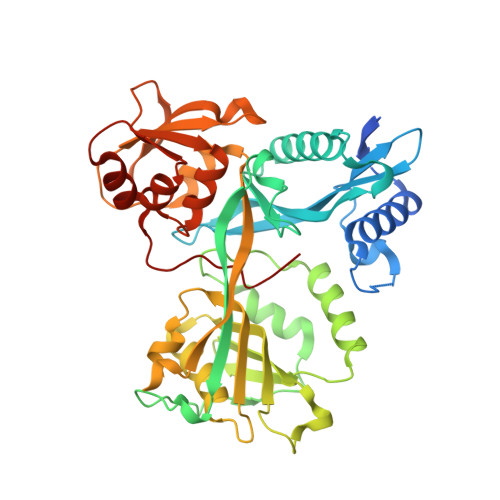
Structural analysis of the N-acetyltransferase Eis1 from Mycobacterium abscessus reveals the molecular determinants of its incapacity to modify aminoglycosides.

Ung, K.L.Kremer, L.Blaise, M.

(2021) Proteins 89: 94-106

  • DOI: https://doi.org/10.1002/prot.25997
  • Primary Citation of Related Structures:  
    6YCA

  • PubMed Abstract: 

    Enhanced intracellular survival (Eis) proteins belonging to the superfamily of the GCN5-related N-acetyltransferases play important functions in mycobacterial pathogenesis. In Mycobacterium tuberculosis, Eis enhances the intracellular survival of the bacilli in macrophages by modulating the host immune response and is capable to chemically modify and inactivate aminoglycosides. In nontuberculous mycobacteria (NTM), Eis shares similar functions. However, Mycobacterium abscessus, a multidrug resistant NTM, possesses two functionally distinct Eis homologues, Eis1 Mab and Eis2 Mab . While Eis2 Mab participates in virulence and aminoglycosides resistance, this is not the case for Eis1 Mab, whose exact biological function remains to be determined. Herein, we show that overexpression of Eis1 Mab in M. abscessus fails to induce resistance to aminoglycosides. To clarify why Eis1 Mab is unable to modify this class of antibiotics, we solved its crystal structure bound to its cofactor, acetyl-CoA. The structure revealed that Eis1 Mab has a typical homohexameric Eis-like organization. The structural analysis supported by biochemical approaches demonstrated that while Eis1 Mab can acetylate small substrates, its active site is too narrow to accommodate aminoglycosides. Comparison with other Eis structures showed that an extended loop between strands 9 and 10 is blocking the access of large substrates to the active site and movement of helices 4 and 5 reduces the volume of the substrate-binding pocket to these compounds in Eis1 Mab . Overall, this study underscores the molecular determinants explaining functional differences between Eis1 Mab and Eis2 Mab, especially those inherent to their capacity to modify aminoglycosides.


  • Organizational Affiliation

    Institut de Recherche en Infectiologie de Montpellier (IRIM), Université de Montpellier, CNRS UMR, Montpellier, France.


Macromolecules
Find similar proteins by:  (by identity cutoff)  |  3D Structure
Entity ID: 1
MoleculeChains Sequence LengthOrganismDetailsImage
Uncharacterized N-acetyltransferase D2E76_00625
A, B, C, D, E
A, B, C, D, E, F
415Mycobacteroides abscessusMutation(s): 0 
Gene Names: D2E76_00625
EC: 2.3.1
UniProt
Find proteins for A0A418LDP6 (Mycobacteroides abscessus)
Explore A0A418LDP6 
Go to UniProtKB:  A0A418LDP6
Entity Groups  
Sequence Clusters30% Identity50% Identity70% Identity90% Identity95% Identity100% Identity
UniProt GroupA0A418LDP6
Sequence Annotations
Expand
  • Reference Sequence
Small Molecules
Ligands 2 Unique
IDChains Name / Formula / InChI Key2D Diagram3D Interactions
ACO (Subject of Investigation/LOI)
Query on ACO

Download Ideal Coordinates CCD File 
CA [auth E]
G [auth A]
GA [auth F]
M [auth B]
T [auth C]
CA [auth E],
G [auth A],
GA [auth F],
M [auth B],
T [auth C],
Z [auth D]
ACETYL COENZYME *A
C23 H38 N7 O17 P3 S
ZSLZBFCDCINBPY-ZSJPKINUSA-N
SO4
Query on SO4

Download Ideal Coordinates CCD File 
AA [auth D]
BA [auth D]
DA [auth E]
EA [auth E]
FA [auth E]
AA [auth D],
BA [auth D],
DA [auth E],
EA [auth E],
FA [auth E],
H [auth A],
HA [auth F],
I [auth A],
IA [auth F],
J [auth A],
K [auth A],
L [auth A],
N [auth B],
O [auth B],
P [auth B],
Q [auth B],
R [auth B],
S [auth B],
U [auth C],
V [auth C],
W [auth C],
X [auth C],
Y [auth C]
SULFATE ION
O4 S
QAOWNCQODCNURD-UHFFFAOYSA-L
Experimental Data & Validation

Experimental Data

  • Method: X-RAY DIFFRACTION
  • Resolution: 2.90 Å
  • R-Value Free: 0.248 
  • R-Value Work: 0.208 
  • R-Value Observed: 0.210 
  • Space Group: P 21 21 21
Unit Cell:
Length ( Å )Angle ( ˚ )
a = 118.39α = 90
b = 150.69β = 90
c = 166.74γ = 90
Software Package:
Software NamePurpose
PHENIXrefinement
PHENIXrefinement
XDSdata reduction
XSCALEdata scaling
MOLREPphasing

Structure Validation

View Full Validation Report


Ligand Structure Quality Assessment 


Entry History 

Deposition Data

Revision History  (Full details and data files)

  • Version 1.0: 2020-09-09
    Type: Initial release
  • Version 1.1: 2020-09-16
    Changes: Database references
  • Version 1.2: 2022-12-21
    Changes: Database references
  • Version 1.3: 2024-01-31
    Changes: Data collection, Refinement description