6YA6

Minimal construct of Cdc7-Dbf4 bound to XL413


Experimental Data Snapshot

  • Method: X-RAY DIFFRACTION
  • Resolution: 1.44 Å
  • R-Value Free: 0.193 
  • R-Value Work: 0.167 
  • R-Value Observed: 0.168 

wwPDB Validation   3D Report Full Report


Ligand Structure Quality Assessment 


This is version 1.3 of the entry. See complete history


Literature

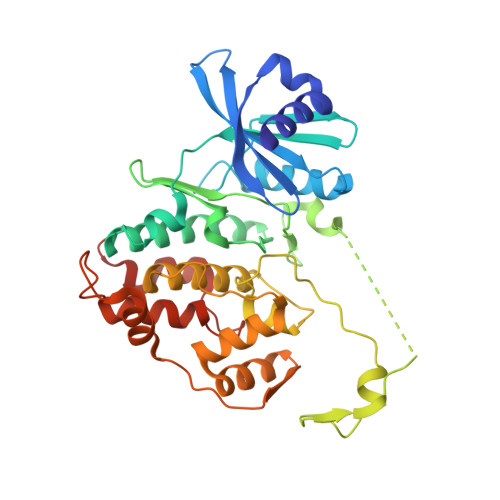
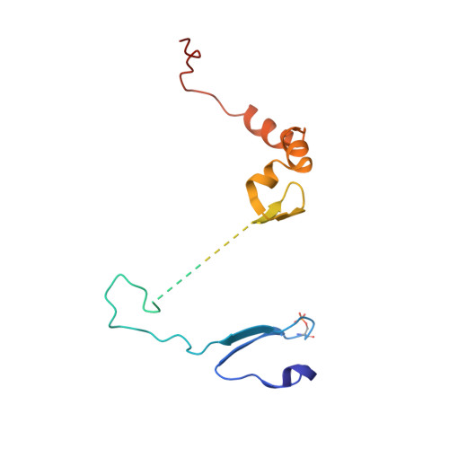
Structural Basis for the Activation and Target Site Specificity of CDC7 Kinase.

Dick, S.D.Federico, S.Hughes, S.M.Pye, V.E.O'Reilly, N.Cherepanov, P.

(2020) Structure 28: 954-962.e4

  • DOI: https://doi.org/10.1016/j.str.2020.05.010
  • Primary Citation of Related Structures:  
    6YA6, 6YA7, 6YA8

  • PubMed Abstract: 

    CDC7 is an essential Ser/Thr kinase that acts upon the replicative helicase throughout the S phase of the cell cycle and is activated by DBF4. Here, we present crystal structures of a highly active human CDC7-DBF4 construct. The structures reveal a zinc-finger domain at the end of the kinase insert 2 that pins the CDC7 activation loop to motif M of DBF4 and the C lobe of CDC7. These interactions lead to ordering of the substrate-binding platform and full opening of the kinase active site. In a co-crystal structure with a mimic of MCM2 Ser40 phosphorylation target, the invariant CDC7 residues Arg373 and Arg380 engage phospho-Ser41 at substrate P+1 position, explaining the selectivity of the S-phase kinase for Ser/Thr residues followed by a pre-phosphorylated or an acidic residue. Our results clarify the role of DBF4 in activation of CDC7 and elucidate the structural basis for recognition of its preferred substrates.


  • Organizational Affiliation

    Chromatin Structure and Mobile DNA, The Francis Crick Institute, London NW1 1AT, UK.


Macromolecules
Find similar proteins by:  (by identity cutoff)  |  3D Structure
Entity ID: 1
MoleculeChains Sequence LengthOrganismDetailsImage
Cell division cycle 7-related protein kinase,Cell division cycle 7-related protein kinase,Cell division cycle 7-related protein kinase359Homo sapiensMutation(s): 0 
Gene Names: CDC7CDC7L1
EC: 2.7.11.1
UniProt & NIH Common Fund Data Resources
Find proteins for O00311 (Homo sapiens)
Explore O00311 
Go to UniProtKB:  O00311
PHAROS:  O00311
GTEx:  ENSG00000097046 
Entity Groups  
Sequence Clusters30% Identity50% Identity70% Identity90% Identity95% Identity100% Identity
UniProt GroupO00311
Sequence Annotations
Expand
  • Reference Sequence
Find similar proteins by:  (by identity cutoff)  |  3D Structure
Entity ID: 2
MoleculeChains Sequence LengthOrganismDetailsImage
Protein DBF4 homolog A144Homo sapiensMutation(s): 0 
Gene Names: DBF4ASKDBF4AZDBF1
UniProt & NIH Common Fund Data Resources
Find proteins for Q9UBU7 (Homo sapiens)
Explore Q9UBU7 
Go to UniProtKB:  Q9UBU7
PHAROS:  Q9UBU7
GTEx:  ENSG00000006634 
Entity Groups  
Sequence Clusters30% Identity50% Identity70% Identity90% Identity95% Identity100% Identity
UniProt GroupQ9UBU7
Sequence Annotations
Expand
  • Reference Sequence
Small Molecules
Ligands 6 Unique
IDChains Name / Formula / InChI Key2D Diagram3D Interactions
0SX (Subject of Investigation/LOI)
Query on 0SX

Download Ideal Coordinates CCD File 
C [auth A]8-chloro-2-[(2S)-pyrrolidin-2-yl][1]benzofuro[3,2-d]pyrimidin-4(3H)-one
C14 H12 Cl N3 O2
JJWLXRKVUJDJKG-VIFPVBQESA-N
PEG
Query on PEG

Download Ideal Coordinates CCD File 
E [auth A]DI(HYDROXYETHYL)ETHER
C4 H10 O3
MTHSVFCYNBDYFN-UHFFFAOYSA-N
IMD
Query on IMD

Download Ideal Coordinates CCD File 
I [auth B]IMIDAZOLE
C3 H5 N2
RAXXELZNTBOGNW-UHFFFAOYSA-O
ZN
Query on ZN

Download Ideal Coordinates CCD File 
D [auth A],
G [auth B]
ZINC ION
Zn
PTFCDOFLOPIGGS-UHFFFAOYSA-N
BO3
Query on BO3

Download Ideal Coordinates CCD File 
F [auth A]BORIC ACID
B H3 O3
KGBXLFKZBHKPEV-UHFFFAOYSA-N
CL
Query on CL

Download Ideal Coordinates CCD File 
H [auth B]CHLORIDE ION
Cl
VEXZGXHMUGYJMC-UHFFFAOYSA-M
Modified Residues  1 Unique
IDChains TypeFormula2D DiagramParent
SEP
Query on SEP
B
L-PEPTIDE LINKINGC3 H8 N O6 PSER
Experimental Data & Validation

Experimental Data

  • Method: X-RAY DIFFRACTION
  • Resolution: 1.44 Å
  • R-Value Free: 0.193 
  • R-Value Work: 0.167 
  • R-Value Observed: 0.168 
  • Space Group: P 41 21 2
Unit Cell:
Length ( Å )Angle ( ˚ )
a = 61.64α = 90
b = 61.64β = 90
c = 233.91γ = 90
Software Package:
Software NamePurpose
PHENIXrefinement
PHENIXrefinement
XDSdata reduction
Aimlessdata scaling
PHASERphasing

Structure Validation

View Full Validation Report


Ligand Structure Quality Assessment 


Entry History & Funding Information

Deposition Data


Funding OrganizationLocationGrant Number
The Francis Crick InstituteUnited KingdomFC10061

Revision History  (Full details and data files)

  • Version 1.0: 2020-05-27
    Type: Initial release
  • Version 1.1: 2020-07-01
    Changes: Database references
  • Version 1.2: 2020-08-19
    Changes: Database references, Derived calculations
  • Version 1.3: 2024-01-24
    Changes: Data collection, Database references, Refinement description