6Y9E

Crystal structure of putative ancestral haloalkane dehalogenase AncHLD2 (node 2)


Experimental Data Snapshot

  • Method: X-RAY DIFFRACTION
  • Resolution: 1.70 Å
  • R-Value Free: 0.218 
  • R-Value Work: 0.181 
  • R-Value Observed: 0.183 

wwPDB Validation   3D Report Full Report


Ligand Structure Quality Assessment 


This is version 1.1 of the entry. See complete history


Literature

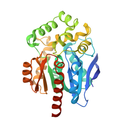
Structures of hyperstable ancestral haloalkane dehalogenases show restricted conformational dynamics.

Babkova, P.Dunajova, Z.Chaloupkova, R.Damborsky, J.Bednar, D.Marek, M.

(2020) Comput Struct Biotechnol J 18: 1497-1508

  • DOI: https://doi.org/10.1016/j.csbj.2020.06.021
  • Primary Citation of Related Structures:  
    6Y9E, 6Y9F, 6Y9G

  • PubMed Abstract: 

    Ancestral sequence reconstruction is a powerful method for inferring ancestors of modern enzymes and for studying structure-function relationships of enzymes. We have previously applied this approach to haloalkane dehalogenases (HLDs) from the subfamily HLD-II and obtained thermodynamically highly stabilized enzymes (Δ T m up to 24 °C), showing improved catalytic properties. Here we combined crystallographic structural analysis and computational molecular dynamics simulations to gain insight into the mechanisms by which ancestral HLDs became more robust enzymes with novel catalytic properties. Reconstructed ancestors exhibited similar structure topology as their descendants with the exception of a few loop deviations. Strikingly, molecular dynamics simulations revealed restricted conformational dynamics of ancestral enzymes, which prefer a single state, in contrast to modern enzymes adopting two different conformational states. The restricted dynamics can potentially be linked to their exceptional stabilization. The study provides molecular insights into protein stabilization due to ancestral sequence reconstruction, which is becoming a widely used approach for obtaining robust protein catalysts.


  • Organizational Affiliation

    Loschmidt Laboratories, Department of Experimental Biology and RECETOX, Faculty of Science, Masaryk University, Kamenice 5, Bld. A13, 625 00 Brno, Czech Republic.


Macromolecules
Find similar proteins by:  (by identity cutoff)  |  3D Structure
Entity ID: 1
MoleculeChains Sequence LengthOrganismDetailsImage
Ancestral haloalkane dehalogenase AncHLD2
A, B, C, D, E
A, B, C, D, E, F
307synthetic constructMutation(s): 0 
Entity Groups  
Sequence Clusters30% Identity50% Identity70% Identity90% Identity95% Identity100% Identity
Sequence Annotations
Expand
  • Reference Sequence
Small Molecules
Ligands 4 Unique
IDChains Name / Formula / InChI Key2D Diagram3D Interactions
TRS (Subject of Investigation/LOI)
Query on TRS

Download Ideal Coordinates CCD File 
J [auth A],
K [auth A],
L [auth A],
M [auth A],
X [auth B]
2-AMINO-2-HYDROXYMETHYL-PROPANE-1,3-DIOL
C4 H12 N O3
LENZDBCJOHFCAS-UHFFFAOYSA-O
MPD (Subject of Investigation/LOI)
Query on MPD

Download Ideal Coordinates CCD File 
HA [auth C],
I [auth A],
IA [auth C],
SA [auth D],
W [auth B]
(4S)-2-METHYL-2,4-PENTANEDIOL
C6 H14 O2
SVTBMSDMJJWYQN-YFKPBYRVSA-N
EDO (Subject of Investigation/LOI)
Query on EDO

Download Ideal Coordinates CCD File 
AA [auth B]
BA [auth B]
BB [auth F]
CA [auth B]
CB [auth F]
AA [auth B],
BA [auth B],
BB [auth F],
CA [auth B],
CB [auth F],
DA [auth B],
DB [auth F],
EA [auth B],
EB [auth F],
FB [auth F],
JA [auth C],
KA [auth C],
LA [auth C],
MA [auth C],
N [auth A],
NA [auth C],
O [auth A],
OA [auth C],
P [auth A],
PA [auth C],
Q [auth A],
R [auth A],
S [auth A],
T [auth A],
TA [auth D],
UA [auth D],
VA [auth D],
Y [auth B],
YA [auth E],
Z [auth B]
1,2-ETHANEDIOL
C2 H6 O2
LYCAIKOWRPUZTN-UHFFFAOYSA-N
CL (Subject of Investigation/LOI)
Query on CL

Download Ideal Coordinates CCD File 
AB [auth F]
FA [auth C]
G [auth A]
GA [auth C]
H [auth A]
AB [auth F],
FA [auth C],
G [auth A],
GA [auth C],
H [auth A],
QA [auth D],
RA [auth D],
U [auth B],
V [auth B],
WA [auth E],
XA [auth E],
ZA [auth F]
CHLORIDE ION
Cl
VEXZGXHMUGYJMC-UHFFFAOYSA-M
Experimental Data & Validation

Experimental Data

  • Method: X-RAY DIFFRACTION
  • Resolution: 1.70 Å
  • R-Value Free: 0.218 
  • R-Value Work: 0.181 
  • R-Value Observed: 0.183 
  • Space Group: P 21 21 2
Unit Cell:
Length ( Å )Angle ( ˚ )
a = 109.58α = 90
b = 166.62β = 90
c = 100.29γ = 90
Software Package:
Software NamePurpose
XDSdata reduction
xia2data scaling
PHASERphasing
PHENIXrefinement
PDB_EXTRACTdata extraction

Structure Validation

View Full Validation Report


Ligand Structure Quality Assessment 


Entry History & Funding Information

Deposition Data


Funding OrganizationLocationGrant Number
European CommissionCzech RepublicMSCA-IF-2017 792772

Revision History  (Full details and data files)

  • Version 1.0: 2020-11-18
    Type: Initial release
  • Version 1.1: 2024-01-24
    Changes: Advisory, Data collection, Database references, Refinement description