6Y6B

Crystal structure of human 14-3-3 gamma in complex with CaMKK2 14-3-3 binding motif Ser100 and 16-OMe-Fusicoccin H


Experimental Data Snapshot

  • Method: X-RAY DIFFRACTION
  • Resolution: 3.08 Å
  • R-Value Free: 0.282 
  • R-Value Work: 0.242 
  • R-Value Observed: 0.244 

wwPDB Validation   3D Report Full Report


Ligand Structure Quality Assessment 


This is version 1.1 of the entry. See complete history


Literature

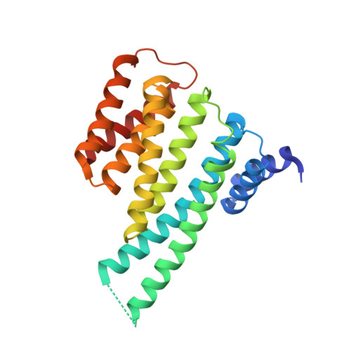
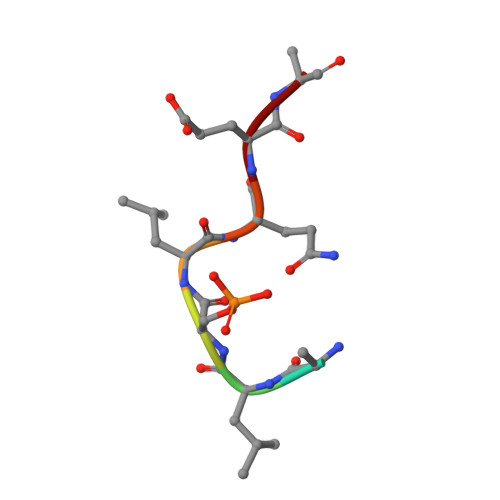
Stabilization of Protein-Protein Interactions between CaMKK2 and 14-3-3 by Fusicoccins.

Lentini Santo, D.Petrvalska, O.Obsilova, V.Ottmann, C.Obsil, T.

(2020) ACS Chem Biol 15: 3060-3071

  • DOI: https://doi.org/10.1021/acschembio.0c00821
  • Primary Citation of Related Structures:  
    6Y4K, 6Y6B

  • PubMed Abstract: 

    Ca 2+ /calmodulin-dependent protein kinase kinase 2 (CaMKK2) regulates several key physiological and pathophysiological processes, and its dysregulation has been implicated in obesity, diabetes, and cancer. CaMKK2 is inhibited through phosphorylation in a process involving binding to the scaffolding 14-3-3 protein, which maintains CaMKK2 in the phosphorylation-mediated inhibited state. The previously reported structure of the N-terminal CaMKK2 14-3-3-binding motif bound to 14-3-3 suggested that the interaction between 14-3-3 and CaMKK2 could be stabilized by small-molecule compounds. Thus, we investigated the stabilization of interactions between CaMKK2 and 14-3-3γ by Fusicoccin A and other fusicoccanes-diterpene glycosides that bind at the interface between the 14-3-3 ligand binding groove and the 14-3-3 binding motif of the client protein. Our data reveal that two of five tested fusicoccanes considerably increase the binding of phosphopeptide representing the 14-3-3 binding motif of CaMKK2 to 14-3-3γ. Crystal structures of two ternary complexes suggest that the steric contacts between the C-terminal part of the CaMKK2 14-3-3 binding motif and the adjacent fusicoccane molecule are responsible for differences in stabilization potency between the study compounds. Moreover, our data also show that fusicoccanes enhance the binding affinity of phosphorylated full-length CaMKK2 to 14-3-3γ, which in turn slows down CaMKK2 dephosphorylation, thus keeping this protein in its phosphorylation-mediated inhibited state. Therefore, targeting the fusicoccin binding cavity of 14-3-3 by small-molecule compounds may offer an alternative strategy to suppress CaMKK2 activity by stabilizing its phosphorylation-mediated inhibited state.


  • Organizational Affiliation

    Department of Physical and Macromolecular Chemistry, Faculty of Science, Charles University, Prague, Czech Republic.


Macromolecules
Find similar proteins by:  (by identity cutoff)  |  3D Structure
Entity ID: 1
MoleculeChains Sequence LengthOrganismDetailsImage
14-3-3 protein gamma
A, B
236Homo sapiensMutation(s): 0 
Gene Names: YWHAG
UniProt & NIH Common Fund Data Resources
Find proteins for P61981 (Homo sapiens)
Explore P61981 
Go to UniProtKB:  P61981
PHAROS:  P61981
GTEx:  ENSG00000170027 
Entity Groups  
Sequence Clusters30% Identity50% Identity70% Identity90% Identity95% Identity100% Identity
UniProt GroupP61981
Sequence Annotations
Expand
  • Reference Sequence

Find similar proteins by:  Sequence   |   3D Structure  

Entity ID: 2
MoleculeChains Sequence LengthOrganismDetailsImage
Calcium/calmodulin-dependent protein kinase kinase 2
C, D
8Homo sapiensMutation(s): 0 
EC: 2.7.11.17
UniProt & NIH Common Fund Data Resources
Find proteins for Q96RR4 (Homo sapiens)
Explore Q96RR4 
Go to UniProtKB:  Q96RR4
PHAROS:  Q96RR4
GTEx:  ENSG00000110931 
Entity Groups  
UniProt GroupQ96RR4
Sequence Annotations
Expand
  • Reference Sequence
Small Molecules
Ligands 1 Unique
IDChains Name / Formula / InChI Key2D Diagram3D Interactions
OD8
Query on OD8

Download Ideal Coordinates CCD File 
E [auth B](2~{R},3~{S},4~{S},5~{R},6~{S})-2-(hydroxymethyl)-6-[[(1~{S},3~{R},6~{S},7~{S},8~{R},9~{R},10~{R},11~{R},14~{S})-14-(me thoxymethyl)-3,10-dimethyl-9-oxidanyl-6-propan-2-yl-8-tricyclo[9.3.0.0^{3,7}]tetradecanyl]oxy]oxane-3,4,5-triol
C27 H48 O8
VEMJQLCDXCMEJD-YTTKSRRZSA-N
Modified Residues  1 Unique
IDChains TypeFormula2D DiagramParent
SEP
Query on SEP
C, D
L-PEPTIDE LINKINGC3 H8 N O6 PSER
Experimental Data & Validation

Experimental Data

  • Method: X-RAY DIFFRACTION
  • Resolution: 3.08 Å
  • R-Value Free: 0.282 
  • R-Value Work: 0.242 
  • R-Value Observed: 0.244 
  • Space Group: P 63 2 2
Unit Cell:
Length ( Å )Angle ( ˚ )
a = 226.266α = 90
b = 226.266β = 90
c = 72.993γ = 120
Software Package:
Software NamePurpose
PHENIXrefinement
XDSdata reduction
XSCALEdata scaling
MOLREPphasing

Structure Validation

View Full Validation Report


Ligand Structure Quality Assessment 


Entry History & Funding Information

Deposition Data


Funding OrganizationLocationGrant Number
Czech Science FoundationCzech Republic19-00121S
European CommissionCzech RepublicTASPPI project, Grant Agreement 675179

Revision History  (Full details and data files)

  • Version 1.0: 2021-02-03
    Type: Initial release
  • Version 1.1: 2024-01-24
    Changes: Data collection, Database references, Refinement description