6Y54 | pdb_00006y54

Crystal structure of a Neisseria meningitidis serogroup A capsular oligosaccharide bound to a functional Fab


Experimental Data Snapshot

  • Method: X-RAY DIFFRACTION
  • Resolution: 2.67 Å
  • R-Value Free: 
    0.267 (Depositor), 0.270 (DCC) 
  • R-Value Work: 
    0.210 (Depositor), 0.212 (DCC) 
  • R-Value Observed: 
    0.213 (Depositor) 

Starting Models: experimental
View more details

wwPDB Validation 3D Report Full Report

Validation slider image for 6Y54

Ligand Structure Quality Assessment 


This is version 1.4 of the entry. See complete history

Literature

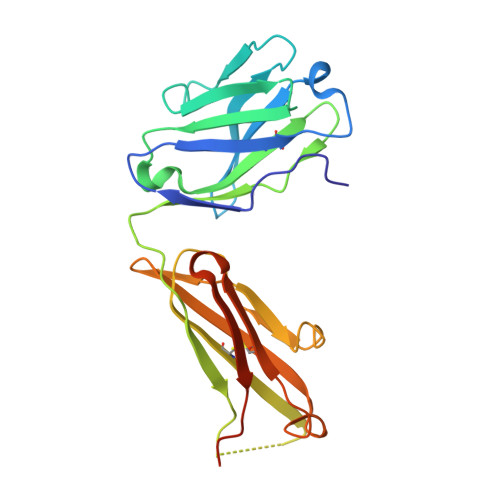
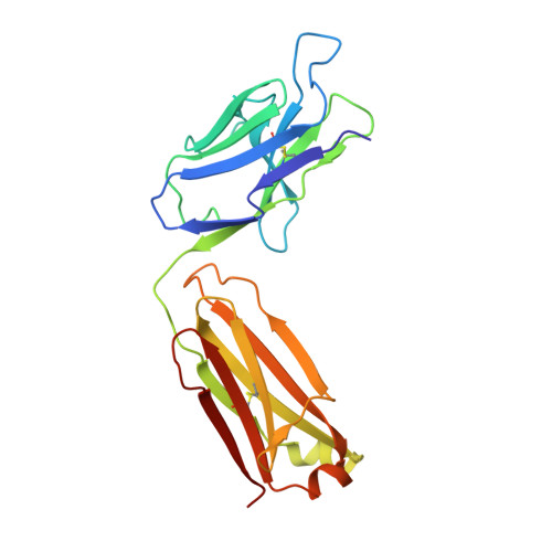
Structure of a protective epitope reveals the importance of acetylation of Neisseria meningitidis serogroup A capsular polysaccharide.

Henriques, P.Dello Iacono, L.Gimeno, A.Biolchi, A.Romano, M.R.Arda, A.Bernardes, G.J.L.Jimenez-Barbero, J.Berti, F.Rappuoli, R.Adamo, R.

(2020) Proc Natl Acad Sci U S A 117: 29795-29802

  • DOI: https://doi.org/10.1073/pnas.2011385117
  • Primary Citation Related Structures: 
    6Y54

  • PubMed Abstract: 

    Meningococcal meningitis remains a substantial cause of mortality and morbidity worldwide. Until recently, countries in the African meningitis belt were susceptible to devastating outbreaks, largely attributed to serogroup A Neisseria meningitidis (MenA). Vaccination with glycoconjugates of MenA capsular polysaccharide led to an almost complete elimination of MenA clinical cases. To understand the molecular basis of vaccine-induced protection, we generated a panel of oligosaccharide fragments of different lengths and tested them with polyclonal and monoclonal antibodies by inhibition enzyme-linked immunosorbent assay, surface plasmon resonance, and competitive human serum bactericidal assay, which is a surrogate for protection. The epitope was shown to optimize between three and six repeating units and to be O -acetylated. The molecular interactions between a protective monoclonal antibody and a MenA capsular polysaccharide fragment were further elucidated at the atomic level by saturation transfer difference NMR spectroscopy and X-ray crystallography. The epitope consists of a trisaccharide anchored to the antibody via the O - and N -acetyl moieties through either H-bonding or CH-π interactions. In silico docking showed that 3- O -acetylation of the upstream residue is essential for antibody binding, while O -acetate could be equally accommodated at three and four positions of the other two residues. These results shed light on the mechanism of action of current MenA vaccines and provide a foundation for the rational design of improved therapies.


  • Organizational Affiliation
    • Instituto de Medicina Molecular João Lobo Antunes, Faculdade de Medicina, Universidade de Lisboa, 1649-028 Lisboa, Portugal.

Macromolecule Content 

  • Total Structure Weight: 150.9 kDa 
  • Atom Count: 8,476 
  • Modeled Residue Count: 1,066 
  • Deposited Residue Count: 1,335 
  • Unique protein chains: 2

Macromolecules

Find similar proteins by:|  3D Structure
Entity ID: 1
MoleculeChains  Sequence LengthOrganismDetailsImage
Fab A1.1 H chainA [auth H],
C [auth M],
E [auth O]
226Mus musculusMutation(s): 0 
Find similar proteins by:|  3D Structure
Entity ID: 2
MoleculeChains  Sequence LengthOrganismDetailsImage
Fab A1.1 L chainB [auth L],
D [auth N],
F [auth P]
219Mus musculusMutation(s): 0 

Small Molecules

Ligands 5 Unique
IDChains Name / Formula / InChI Key2D Diagram3D Interactions
OOW

Query on OOW


Download:Ideal Coordinates CCD File
K [auth H],
U [auth M],
Y [auth O]
{[(2R,3S,4R,5S,6R)-5-acetamido-3-hydroxy-4-(2-oxopropyl)-6-(phosphonooxy)oxan-2-yl]methoxy}phosphonic acid
C10 H19 N O13 P2
WZRFFNAOXAJBLK-JDDHQFAOSA-N
OAB

Query on OAB


Download:Ideal Coordinates CCD File
AA [auth P],
O [auth L],
X [auth N]
{[(2R,3S,4R,5S,6S)-5-acetamido-3,6-dihydroxy-4-(2-oxopropyl)oxan-2-yl]methoxy}phosphonic acid
C10 H18 N O10 P
LMXWGHPJIXYEIE-QWIPYHCPSA-N
OA8

Query on OA8


Download:Ideal Coordinates CCD File
N [auth L],
W [auth N],
Z [auth P]
{[(2R,3S,4R,5S,6S)-3-(acetyloxy)-5-acetamido-4,6-dihydroxyoxan-2-yl]methoxy}phosphonic acid
C10 H18 N O10 P
WZFGOBLYIBEZRF-QWIPYHCPSA-N
IOD

Query on IOD


Download:Ideal Coordinates CCD File
G [auth H]
H
L
M [auth L]
P [auth M]
G [auth H],
H,
L,
M [auth L],
P [auth M],
Q [auth M],
R [auth M],
S [auth M],
V [auth N]
IODIDE ION
I
XMBWDFGMSWQBCA-UHFFFAOYSA-M
EDO

Query on EDO


Download:Ideal Coordinates CCD File
I [auth H],
J [auth H],
T [auth M]
1,2-ETHANEDIOL
C2 H6 O2
LYCAIKOWRPUZTN-UHFFFAOYSA-N

Experimental Data & Validation

Experimental Data

  • Method: X-RAY DIFFRACTION
  • Resolution: 2.67 Å
  • R-Value Free:  0.267 (Depositor), 0.270 (DCC) 
  • R-Value Work:  0.210 (Depositor), 0.212 (DCC) 
  • R-Value Observed: 0.213 (Depositor) 
Space Group: P 1 21 1
Unit Cell:
Length ( Å )Angle ( ˚ )
a = 52.609α = 90
b = 153.205β = 91.527
c = 101.051γ = 90
Software Package:
Software NamePurpose
PHENIXrefinement
XDSdata reduction
Aimlessdata scaling
MOLREPphasing

Structure Validation

View Full Validation Report


Ligand Structure Quality Assessment 


Entry History 

& Funding Information

Deposition Data


Funding OrganizationLocationGrant Number
European CommissionItaly675671

Revision History  (Full details and data files)

  • Version 1.0: 2020-10-07
    Type: Initial release
  • Version 1.1: 2020-11-18
    Changes: Database references
  • Version 1.2: 2020-12-02
    Changes: Database references
  • Version 1.3: 2024-01-24
    Changes: Advisory, Data collection, Database references, Refinement description
  • Version 1.4: 2024-10-09
    Changes: Structure summary