6Y3Q | pdb_00006y3q

Streptavidin mutant S112R_K121E with a biotC5-1 cofactor - an artificial iron hydroxylase


Experimental Data Snapshot

  • Method: X-RAY DIFFRACTION
  • Resolution: 1.95 Å
  • R-Value Free: 
    0.258 (Depositor), 0.257 (DCC) 
  • R-Value Work: 
    0.209 (Depositor), 0.209 (DCC) 

Starting Model: experimental
View more details

wwPDB Validation 3D Report Full Report

Validation slider image for 6Y3Q

Ligand Structure Quality Assessment 


This is version 1.1 of the entry. See complete history

Literature

Enantioselective Hydroxylation of Benzylic C(sp3)-H Bonds by an Artificial Iron Hydroxylase Based on the Biotin-Streptavidin Technology.

Serrano-Plana, J.Rumo, C.Rebelein, J.G.Peterson, R.L.Barnet, M.Ward, T.R.

(2020) J Am Chem Soc 142: 10617-10623

  • DOI: https://doi.org/10.1021/jacs.0c02788
  • Primary Citation Related Structures: 
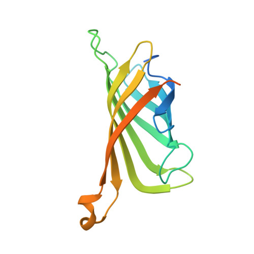
    6Y25, 6Y2M, 6Y2T, 6Y33, 6Y34, 6Y3Q

  • PubMed Abstract: 

    The selective hydroxylation of C-H bonds is of great interest to the synthetic community. Both homogeneous catalysts and enzymes offer complementary means to tackle this challenge. Herein, we show that biotinylated Fe(TAML)-complexes (TAML = Tetra Amido Macrocyclic Ligand) can be used as cofactors for incorporation into streptavidin to assemble artificial hydroxylases. Chemo-genetic optimization of both cofactor and streptavidin allowed optimizing the performance of the hydroxylase. Using H 2 O 2 as oxidant, up to ∼300 turnovers for the oxidation of benzylic C-H bonds were obtained. Upgrading the ee was achieved by kinetic resolution of the resulting benzylic alcohol to afford up to >98% ee for ( R )-tetralol. X-ray analysis of artificial hydroxylases highlights critical details of the second coordination sphere around the Fe(TAML) cofactor.


  • Organizational Affiliation
    • Department of Chemistry, University of Basel, BPR1096, Mattenstrasse 24a, CH-4058 Basel, Switzerland.

Macromolecule Content 

  • Total Structure Weight: 17.42 kDa 
  • Atom Count: 995 
  • Modeled Residue Count: 120 
  • Deposited Residue Count: 159 
  • Unique protein chains: 1

Macromolecules

Find similar proteins by:|  3D Structure
Entity ID: 1
MoleculeChains  Sequence LengthOrganismDetailsImage
StreptavidinA [auth AAA]159Streptomyces avidiniiMutation(s): 2 
UniProt
Find proteins for P22629 (Streptomyces avidinii)
Explore P22629 
Go to UniProtKB:  P22629
Entity Groups
Sequence Clusters30% Identity50% Identity70% Identity90% Identity95% Identity100% Identity
UniProt GroupP22629
Sequence Annotations
Expand
Reference Sequence

Experimental Data & Validation

Experimental Data

  • Method: X-RAY DIFFRACTION
  • Resolution: 1.95 Å
  • R-Value Free:  0.258 (Depositor), 0.257 (DCC) 
  • R-Value Work:  0.209 (Depositor), 0.209 (DCC) 
Space Group: I 41 2 2
Unit Cell:
Length ( Å )Angle ( ˚ )
a = 57.261α = 90
b = 57.261β = 90
c = 182.104γ = 90
Software Package:
Software NamePurpose
REFMACrefinement
XDSdata reduction
Aimlessdata scaling
PHASERphasing

Structure Validation

View Full Validation Report


Ligand Structure Quality Assessment 


Entry History 

& Funding Information

Deposition Data


Funding OrganizationLocationGrant Number
Swiss National Science FoundationSwitzerland200020_182046
Swiss National Science FoundationSwitzerlandNCCR MSE
European Research Council (ERC)European Union694424
European Molecular Biology Organization (EMBO)GermanyALTF 194-2017
European Research Council (ERC)European UnionH2020-MSCA-IF-2016

Revision History  (Full details and data files)

  • Version 1.0: 2020-07-01
    Type: Initial release
  • Version 1.1: 2024-01-24
    Changes: Data collection, Database references, Derived calculations, Refinement description