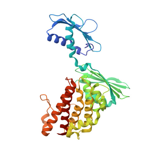
Alternative dimerization is required for activity and inhibition of the HEPN ribonuclease RnlA.
Garcia-Rodriguez, G., Charlier, D., Wilmaerts, D., Michiels, J., Loris, R.(2021) Nucleic Acids Res 49: 7164-7178
- PubMed: 34139012 Search on PubMedSearch on PubMed Central
- DOI: https://doi.org/10.1093/nar/gkab513
- Primary Citation Related Structures:
6Y2P, 6Y2Q, 6Y2R, 7AEX - PubMed Abstract:
The rnlAB toxin-antitoxin operon from Escherichia coli functions as an anti-phage defense system. RnlA was identified as a member of the HEPN (Higher Eukaryotes and Prokaryotes Nucleotide-binding domain) superfamily of ribonucleases. The activity of the toxin RnlA requires tight regulation by the antitoxin RnlB, the mechanism of which remains unknown. Here we show that RnlA exists in an equilibrium between two different homodimer states: an inactive resting state and an active canonical HEPN dimer. Mutants interfering with the transition between states show that canonical HEPN dimerization via the highly conserved RX4-6H motif is required for activity. The antitoxin RnlB binds the canonical HEPN dimer conformation, inhibiting RnlA by blocking access to its active site. Single-alanine substitutions mutants of the highly conserved R255, E258, R318 and H323 show that these residues are involved in catalysis and substrate binding and locate the catalytic site near the dimer interface of the canonical HEPN dimer rather than in a groove located between the HEPN domain and the preceding TBP-like domain. Overall, these findings elucidate the structural basis of the activity and inhibition of RnlA and highlight the crucial role of conformational heterogeneity in protein function.
- Structural Biology Brussels, Department of Biotechnology, Vrije Universiteit Brussel, B-1050 Brussel, Belgium.
Organizational Affiliation: