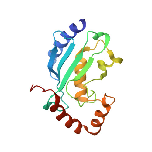
1H,13C,15N backbone and side-chain resonance assignment of the native form of UbcH7 (UBE2L3) through solution NMR spectroscopy.
Marousis, K.D., Birkou, M., Asimakopoulou, A., Spyroulias, G.A.(2020) Biomol NMR Assign 14: 73-78
- PubMed: 31792831
- DOI: https://doi.org/10.1007/s12104-019-09923-9
- Primary Citation of Related Structures:
6XXU - PubMed Abstract:
Ubiquitination is a post-translational modification that regulates a plethora of processes in cells. Ubiquitination requires three type of enzyme: E1 ubiquitin (Ub) activating enzymes, E2 Ub conjugating enzymes and E3 ubiquitin ligases. The E2 enzymes perform a variety of functions, as Ub chain initiation, elongation and regulation of the topology and the process of chain formation. The E2 enzymes family is mainly characterized by a highly conserved ubiquitin conjugating domain (UBC), which comprises the binding region for the activated Ub, E1 and E3 enzymes. The E2 enzyme UbcH7 (UBE2L3) is a known interacting partner for different types of E3 Ub ligases such as HECT, RING and RBR. A structural analysis of the apo form of the native UbcH7 will provide the structural information to understand how this E2 enzyme is implicated in a wide range of diseases and how it interacts with its partners. In the present study we present the high yield expression of the native UbcH7 E2 enzyme and its preliminary analysis via solution NMR spectroscopy. The E2 enzyme is folded in solution and nearly a complete backbone assignment was achieved. Additionally, TALOS+ analysis was performed and the results indicated that UbcH7 adopts a αββββααα topology which is similar to that of the majority of E2 enzymes.
- Department of Pharmacy, University of Patras, 26504, Patras, Greece.
Organizational Affiliation:


