6XWP | pdb_00006xwp

Structure of glutamate transporter homologue GltTk in unsaturated conditions - outward-outward-inward configuration


Experimental Data Snapshot

  • Method: ELECTRON MICROSCOPY
  • Resolution: 3.38 Å
  • Aggregation State: PARTICLE 
  • Reconstruction Method: SINGLE PARTICLE 

wwPDB Validation 3D Report Full Report

Validation slider image for 6XWP

This is version 1.2 of the entry. See complete history

Literature

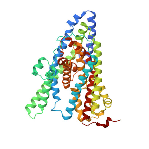
Structural ensemble of a glutamate transporter homologue in lipid nanodisc environment.

Arkhipova, V.Guskov, A.Slotboom, D.J.

(2020) Nat Commun 11: 998-998

  • DOI: https://doi.org/10.1038/s41467-020-14834-8
  • Primary Citation Related Structures: 
    6XWN, 6XWO, 6XWP, 6XWQ, 6XWR

  • PubMed Abstract: 

    Glutamate transporters are cation-coupled secondary active membrane transporters that clear the neurotransmitter L-glutamate from the synaptic cleft. These transporters are homotrimers, with each protomer functioning independently by an elevator-type mechanism, in which a mobile transport domain alternates between inward- and outward-oriented states. Using single-particle cryo-EM we have determined five structures of the glutamate transporter homologue Glt Tk , a Na + - L-aspartate symporter, embedded in lipid nanodiscs. Dependent on the substrate concentrations used, the protomers of the trimer adopt a variety of asymmetrical conformations, consistent with the independent movement. Six of the 15 resolved protomers are in a hitherto elusive state of the transport cycle in which the inward-facing transporters are loaded with Na + ions. These structures explain how substrate-leakage is prevented - a strict requirement for coupled transport. The belt protein of the lipid nanodiscs bends around the inward oriented protomers, suggesting that membrane deformations occur during transport.


  • Organizational Affiliation
    • Groningen Biomolecular Sciences and Biotechnology Institute (GBB), University of Groningen, Nijenborgh 4, 9747AG, Groningen, The Netherlands.

Macromolecule Content 

  • Total Structure Weight: 137.01 kDa 
  • Atom Count: 9,552 
  • Modeled Residue Count: 1,278 
  • Deposited Residue Count: 1,290 
  • Unique protein chains: 1

Macromolecules

Find similar proteins by:|  3D Structure
Entity ID: 1
MoleculeChains  Sequence LengthOrganismDetailsImage
Proton/glutamate symporter, SDF family
A, B, C
430Thermococcus kodakarensis KOD1Mutation(s): 0 
Gene Names: TK0986
Membrane Entity: Yes 
UniProt
Find proteins for Q5JID0 (Thermococcus kodakarensis (strain ATCC BAA-918 / JCM 12380 / KOD1))
Explore Q5JID0 
Go to UniProtKB:  Q5JID0
Entity Groups
Sequence Clusters30% Identity50% Identity70% Identity90% Identity95% Identity100% Identity
UniProt GroupQ5JID0
Sequence Annotations
Expand
Reference Sequence

Experimental Data & Validation

Experimental Data

  • Method: ELECTRON MICROSCOPY
  • Resolution: 3.38 Å
  • Aggregation State: PARTICLE 
  • Reconstruction Method: SINGLE PARTICLE 
EM Software:
TaskSoftware PackageVersion
MODEL REFINEMENTPHENIX

Structure Validation

View Full Validation Report


Entry History 

Deposition Data

Revision History  (Full details and data files)

  • Version 1.0: 2020-03-04
    Type: Initial release
  • Version 1.1: 2024-05-22
    Changes: Data collection, Database references
  • Version 1.2: 2025-07-02
    Changes: Data collection, Structure summary