6XVC | pdb_00006xvc

CRYSTAL STRUCTURE OF BRD4-BD1 WITH COMPOUND 1


Experimental Data Snapshot

  • Method: X-RAY DIFFRACTION
  • Resolution: 1.10 Å
  • R-Value Free: 
    0.198 (Depositor), 0.201 (DCC) 
  • R-Value Work: 
    0.172 (Depositor), 0.177 (DCC) 
  • R-Value Observed: 
    0.173 (Depositor) 

Starting Model: experimental
View more details

wwPDB Validation 3D Report Full Report

Validation slider image for 6XVC

Ligand Structure Quality Assessment 


This is version 1.3 of the entry. See complete history

Literature

PI by NMR: Probing CH-pi Interactions in Protein-Ligand Complexes by NMR Spectroscopy.

Platzer, G.Mayer, M.Beier, A.Bruschweiler, S.Fuchs, J.E.Engelhardt, H.Geist, L.Bader, G.Schorghuber, J.Lichtenecker, R.Wolkerstorfer, B.Kessler, D.McConnell, D.B.Konrat, R.

(2020) Angew Chem Int Ed Engl 59: 14861-14868

  • DOI: https://doi.org/10.1002/anie.202003732
  • Primary Citation Related Structures: 
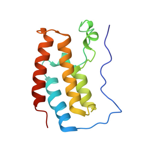
    6XUZ, 6XV3, 6XV7, 6XVC

  • PubMed Abstract: 

    While CH-π interactions with target proteins are crucial determinants for the affinity of arguably every drug molecule, no method exists to directly measure the strength of individual CH-π interactions in drug-protein complexes. Herein, we present a fast and reliable methodology called PI (π interactions) by NMR, which can differentiate the strength of protein-ligand CH-π interactions in solution. By combining selective amino-acid side-chain labeling with 1 H- 13 C NMR, we are able to identify specific protein protons of side-chains engaged in CH-π interactions with aromatic ring systems of a ligand, based solely on 1 H chemical-shift values of the interacting protein aromatic ring protons. The information encoded in the chemical shifts induced by such interactions serves as a proxy for the strength of each individual CH-π interaction. PI by NMR changes the paradigm by which chemists can optimize the potency of drug candidates: direct determination of individual π interactions rather than averaged measures of all interactions.


  • Organizational Affiliation
    • Christian Doppler Laboratory for High-Content Structural Biology and Biotechnology, Department of Structural and Computational Biology, Max Perutz Labs, University of Vienna, Campus Vienna Biocenter 5, 1030, Vienna, Austria.

Macromolecule Content 

  • Total Structure Weight: 15.61 kDa 
  • Atom Count: 1,208 
  • Modeled Residue Count: 125 
  • Deposited Residue Count: 127 
  • Unique protein chains: 1

Macromolecules

Find similar proteins by:|  3D Structure
Entity ID: 1
MoleculeChains  Sequence LengthOrganismDetailsImage
Bromodomain-containing protein 4A [auth B]127Homo sapiensMutation(s): 0 
Gene Names: BRD4HUNK1
UniProt & NIH Common Fund Data Resources
Find proteins for O60885 (Homo sapiens)
Explore O60885 
Go to UniProtKB:  O60885
PHAROS:  O60885
GTEx:  ENSG00000141867 
Entity Groups
Sequence Clusters30% Identity50% Identity70% Identity90% Identity95% Identity100% Identity
UniProt GroupO60885
Sequence Annotations
Expand
Reference Sequence

Small Molecules

Experimental Data & Validation

Experimental Data

  • Method: X-RAY DIFFRACTION
  • Resolution: 1.10 Å
  • R-Value Free:  0.198 (Depositor), 0.201 (DCC) 
  • R-Value Work:  0.172 (Depositor), 0.177 (DCC) 
  • R-Value Observed: 0.173 (Depositor) 
Space Group: P 21 21 21
Unit Cell:
Length ( Å )Angle ( ˚ )
a = 39.197α = 90
b = 50.466β = 90
c = 57.537γ = 90
Software Package:
Software NamePurpose
BUSTERrefinement
XDSdata reduction
autoPROCdata reduction
Aimlessdata scaling
STARANISOdata scaling
PHASERphasing

Structure Validation

View Full Validation Report


Ligand Structure Quality Assessment 


Entry History 

Deposition Data

Revision History  (Full details and data files)

  • Version 1.0: 2020-07-08
    Type: Initial release
  • Version 1.1: 2020-07-22
    Changes: Database references, Refinement description
  • Version 1.2: 2021-03-10
    Changes: Database references
  • Version 1.3: 2024-01-24
    Changes: Data collection, Database references, Refinement description