Biochemical and crystallographic investigations into isonitrile formation by a nonheme iron-dependent oxidase/decarboxylase.
Jonnalagadda, R., Del Rio Flores, A., Cai, W., Mehmood, R., Narayanamoorthy, M., Ren, C., Zaragoza, J.P.T., Kulik, H.J., Zhang, W., Drennan, C.L.(2021) J Biol Chem 296: 100231-100231
- PubMed: 33361191 Search on PubMedSearch on PubMed Central
- DOI: https://doi.org/10.1074/jbc.RA120.015932
- Primary Citation Related Structures:
6XN6, 6XO3, 6XOJ, 6XPA - PubMed Abstract:
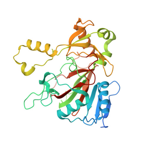
The isonitrile moiety is found in marine sponges and some microbes, where it plays a role in processes such as virulence and metal acquisition. Until recently only one route was known for isonitrile biosynthesis, a condensation reaction that brings together a nitrogen atom of l-Trp/l-Tyr with a carbon atom from ribulose-5-phosphate. With the discovery of ScoE, a mononuclear Fe(II) α-ketoglutarate-dependent dioxygenase from Streptomyces coeruleorubidus, a second route was identified. ScoE forms isonitrile from a glycine adduct, with both the nitrogen and carbon atoms coming from the same glycyl moiety. This reaction is part of the nonribosomal biosynthetic pathway of isonitrile lipopeptides. Here, we present structural, biochemical, and computational investigations of the mechanism of isonitrile formation by ScoE, an unprecedented reaction in the mononuclear Fe(II) α-ketoglutarate-dependent dioxygenase superfamily. The stoichiometry of this enzymatic reaction is measured, and multiple high-resolution (1.45-1.96 Å resolution) crystal structures of Fe(II)-bound ScoE are presented, providing insight into the binding of substrate, (R)-3-((carboxylmethyl)amino)butanoic acid (CABA), cosubstrate α-ketoglutarate, and an Fe(IV)=O mimic oxovanadium. Comparison to a previously published crystal structure of ScoE suggests that ScoE has an "inducible" α-ketoglutarate binding site, in which two residues arginine-157 and histidine-299 move by approximately 10 Å from the surface of the protein into the active site to create a transient α-ketoglutarate binding pocket. Together, data from structural analyses, site-directed mutagenesis, and computation provide insight into the mode of α-ketoglutarate binding, the mechanism of isonitrile formation, and how the structure of ScoE has been adapted to perform this unusual chemical reaction.
- Department of Biology, Massachusetts Institute of Technology, Cambridge, Massachusetts, USA.
Organizational Affiliation: