6XM9

Crystal structure of vanillin bound to Co-LSD4 from Sphingobium sp. strain SYK-6


Experimental Data Snapshot

  • Method: X-RAY DIFFRACTION
  • Resolution: 1.65 Å
  • R-Value Free: 0.187 
  • R-Value Work: 0.159 
  • R-Value Observed: 0.160 

wwPDB Validation   3D Report Full Report


Ligand Structure Quality Assessment 


This is version 1.3 of the entry. See complete history


Literature

Structural and functional analysis of lignostilbene dioxygenases from Sphingobium sp. SYK-6.

Kuatsjah, E.Chan, A.C.K.Katahira, R.Haugen, S.J.Beckham, G.T.Murphy, M.E.P.Eltis, L.D.

(2021) J Biol Chem 296: 100758-100758

  • DOI: https://doi.org/10.1016/j.jbc.2021.100758
  • Primary Citation of Related Structures:  
    6XM6, 6XM7, 6XM8, 6XM9, 6XMA

  • PubMed Abstract: 

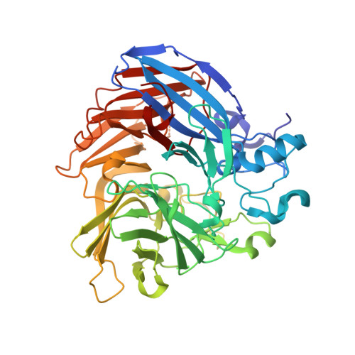
    Lignostilbene-α,β-dioxygenases (LSDs) are iron-dependent oxygenases involved in the catabolism of lignin-derived stilbenes. Sphingobium sp. SYK-6 contains eight LSD homologs with undetermined physiological roles. To investigate which homologs are involved in the catabolism of dehydrodiconiferyl alcohol (DCA), derived from β-5 linked lignin subunits, we heterologously produced the enzymes and screened their activities in lysates. The seven soluble enzymes all cleaved lignostilbene, but only LSD2, LSD3, and LSD4 exhibited high specific activity for 3-(4-hydroxy-3-(4-hydroxy-3-methoxystyryl)-5-methoxyphenyl) acrylate (DCA-S) relative to lignostilbene. LSD4 catalyzed the cleavage of DCA-S to 5-formylferulate and vanillin and cleaved lignostilbene and DCA-S (∼10 6  M -1 s -1 ) with tenfold greater specificity than pterostilbene and resveratrol. X-ray crystal structures of native LSD4 and the catalytically inactive cobalt-substituted Co-LSD4 at 1.45 Å resolution revealed the same fold, metal ion coordination, and edge-to-edge dimeric structure as observed in related enzymes. Key catalytic residues, Phe-59, Tyr-101, and Lys-134, were also conserved. Structures of Co-LSD4·vanillin, Co-LSD4·lignostilbene, and Co-LSD4·DCA-S complexes revealed that Ser-283 forms a hydrogen bond with the hydroxyl group of the ferulyl portion of DCA-S. This residue is conserved in LSD2 and LSD4 but is alanine in LSD3. Substitution of Ser-283 with Ala minimally affected the specificity of LSD4 for either lignostilbene or DCA-S. By contrast, substitution with phenylalanine, as occurs in LSD5 and LSD6, reduced the specificity of the enzyme for both substrates by an order of magnitude. This study expands our understanding of an LSD critical to DCA catabolism as well as the physiological roles of other LSDs and their determinants of substrate specificity.


  • Organizational Affiliation

    Department of Microbiology and Immunology, Life Sciences Institute, The University of British Columbia, Vancouver, Canada; Renewable Resources and Enabling Sciences Center, National Renewable Energy Laboratory, Golden, Colorado, USA; Center for Bioenergy Innovation, Oak Ridge National Laboratory, Oak Ridge Tennessee, USA.


Macromolecules
Find similar proteins by:  (by identity cutoff)  |  3D Structure
Entity ID: 1
MoleculeChains Sequence LengthOrganismDetailsImage
Dioxygenase489Sphingobium sp. SYK-6Mutation(s): 0 
Gene Names: SLG_12860
EC: 1.13.11
UniProt
Find proteins for G2IQT9 (Sphingobium sp. (strain NBRC 103272 / SYK-6))
Explore G2IQT9 
Go to UniProtKB:  G2IQT9
Entity Groups  
Sequence Clusters30% Identity50% Identity70% Identity90% Identity95% Identity100% Identity
UniProt GroupG2IQT9
Sequence Annotations
Expand
  • Reference Sequence
Small Molecules
Ligands 5 Unique
IDChains Name / Formula / InChI Key2D Diagram3D Interactions
V55 (Subject of Investigation/LOI)
Query on V55

Download Ideal Coordinates CCD File 
B [auth A]4-hydroxy-3-methoxybenzaldehyde
C8 H8 O3
MWOOGOJBHIARFG-UHFFFAOYSA-N
DMS
Query on DMS

Download Ideal Coordinates CCD File 
C [auth A]
D [auth A]
E [auth A]
F [auth A]
H [auth A]
C [auth A],
D [auth A],
E [auth A],
F [auth A],
H [auth A],
I [auth A],
J [auth A],
K [auth A]
DIMETHYL SULFOXIDE
C2 H6 O S
IAZDPXIOMUYVGZ-UHFFFAOYSA-N
ACT
Query on ACT

Download Ideal Coordinates CCD File 
G [auth A]ACETATE ION
C2 H3 O2
QTBSBXVTEAMEQO-UHFFFAOYSA-M
CO (Subject of Investigation/LOI)
Query on CO

Download Ideal Coordinates CCD File 
L [auth A],
M [auth A]
COBALT (II) ION
Co
XLJKHNWPARRRJB-UHFFFAOYSA-N
MG
Query on MG

Download Ideal Coordinates CCD File 
N [auth A],
O [auth A]
MAGNESIUM ION
Mg
JLVVSXFLKOJNIY-UHFFFAOYSA-N
Experimental Data & Validation

Experimental Data

  • Method: X-RAY DIFFRACTION
  • Resolution: 1.65 Å
  • R-Value Free: 0.187 
  • R-Value Work: 0.159 
  • R-Value Observed: 0.160 
  • Space Group: I 2 2 2
Unit Cell:
Length ( Å )Angle ( ˚ )
a = 85.16α = 90
b = 112.157β = 90
c = 115.311γ = 90
Software Package:
Software NamePurpose
PHENIXrefinement
DENZOdata reduction
HKL-2000data scaling
PDB_EXTRACTdata extraction
MOLREPphasing

Structure Validation

View Full Validation Report


Ligand Structure Quality Assessment 


Entry History & Funding Information

Deposition Data


Funding OrganizationLocationGrant Number
Natural Sciences and Engineering Research Council (NSERC, Canada)Canada04802
Natural Sciences and Engineering Research Council (NSERC, Canada)Canada171359

Revision History  (Full details and data files)

  • Version 1.0: 2021-05-12
    Type: Initial release
  • Version 1.1: 2021-05-26
    Changes: Database references
  • Version 1.2: 2021-07-21
    Changes: Database references
  • Version 1.3: 2023-10-18
    Changes: Data collection, Database references, Refinement description