Peptidoglycan binding by a pocket on the accessory NTF2-domain of Pgp2 directs helical cell shape of Campylobacter jejuni.
Lin, C.S., Chan, A.C.K., Vermeulen, J., Brockerman, J., Soni, A.S., Tanner, M.E., Gaynor, E.C., McIntosh, L.P., Simorre, J.P., Murphy, M.E.P.(2021) J Biol Chem 296: 100528-100528
- PubMed: 33711341 Search on PubMedSearch on PubMed Central
- DOI: https://doi.org/10.1016/j.jbc.2021.100528
- Primary Citation Related Structures:
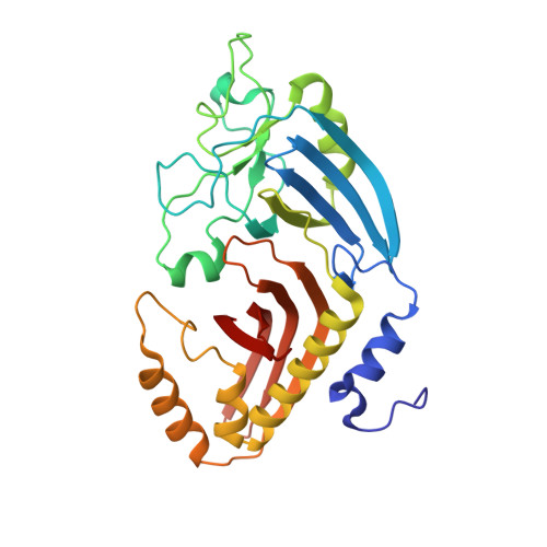
6XJ6, 6XJ7 - PubMed Abstract:
The helical morphology of Campylobacter jejuni, a bacterium involved in host gut colonization and pathogenesis in humans, is determined by the structure of the peptidoglycan (PG) layer. This structure is dictated by trimming of peptide stems by the LD-carboxypeptidase Pgp2 within the periplasm. The interaction interface between Pgp2 and PG to select sites for peptide trimming is unknown. We determined a 1.6 Å resolution crystal structure of Pgp2, which contains a conserved LD-carboxypeptidase domain and a previously uncharacterized domain with an NTF2-like fold (NTF2). We identified a pocket in the NTF2 domain formed by conserved residues and located ∼40 Å from the LD-carboxypeptidase active site. Expression of pgp2 in trans with substitutions of charged (Lys257, Lys307, Glu324) and hydrophobic residues (Phe242 and Tyr233) within the pocket did not restore helical morphology to a pgp2 deletion strain. Muropeptide analysis indicated a decrease of murotripeptides in the deletion strain expressing these mutants, suggesting reduced Pgp2 catalytic activity. Pgp2 but not the K307A mutant was pulled down by C. jejuni Δpgp2 PG sacculi, supporting a role for the pocket in PG binding. NMR spectroscopy was used to define the interaction interfaces of Pgp2 with several PG fragments, which bound to the active site within the LD-carboxypeptidase domain and the pocket of the NTF2 domain. We propose a model for Pgp2 binding to PG strands involving both the LD-carboxypeptidase domain and the accessory NTF2 domain to induce a helical cell shape.
- Department of Microbiology and Immunology, University of British Columbia, Vancouver, British Columbia, Canada.
Organizational Affiliation: